

Heterobivalent agents targeting PSMA and integrin-αvβ3
- PMID: 24410012
- PMCID: PMC4112557
- DOI: 10.1021/bc4005377
Heterobivalent agents targeting PSMA and integrin-αvβ3
Abstract
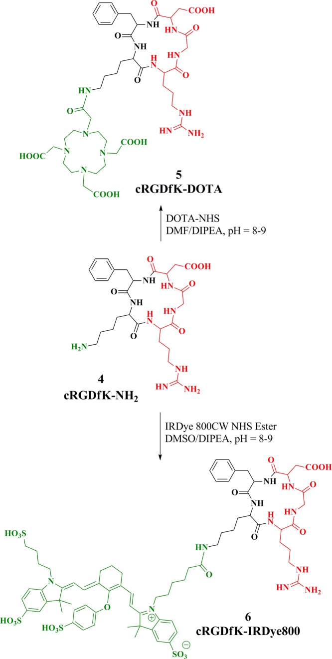
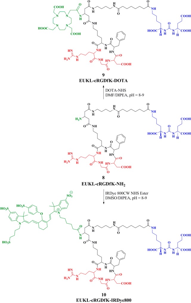
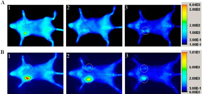
Differential expression of surface proteins on normal vs malignant cells provides the rationale for the development of receptor-, antigen-, and transporter-based, cancer-selective imaging and therapeutic agents. However, tumors are heterogeneous, and do not always express what can be considered reliable, tumor-selective markers. That suggests development of more flexible targeting platforms that incorporate multiple moieties enabling concurrent targeting to a variety of putative markers. We report the synthesis, biochemical, in vitro, and preliminary in vivo evaluation of a new heterobivalent (HtBv) imaging agent targeting both the prostate-specific membrane antigen (PSMA) and integrin-αvβ3 surface markers, each of which can be overexpressed in certain tumor epithelium and/or neovasculature. The HtBv agent was functionalized with either 1,4,7,10-tetraazacyclododecane-1,4,7,10-tetraacetic acid (DOTA) or the commercially available IRDye800CW. DOTA-conjugated HtBv probe 9 bound to PSMA or αvβ3 with affinities similar to those of monovalent (Mnv) compounds designed to bind to their targets independently. In situ energy minimization experiments support a model describing the conformations adapted by 9 that enable it to bind both targets. IRDye800-conjugated HtBv probe 10 demonstrated target-specific binding to either PSMA or integrin-αvβ3 overexpressing xenografts. HtBv agents 9 and 10 may enable dual-targeted imaging of malignant cells and tissues in an effort to address heterogeneity that confounds many cancer-targeted imaging agents.
Figures


Similar articles
-
A Novel ¹¹¹In-Labeled Anti-Prostate-Specific Membrane Antigen Nanobody for Targeted SPECT/CT Imaging of Prostate Cancer.J Nucl Med. 2015 Jul;56(7):1094-9. doi: 10.2967/jnumed.115.156729. Epub 2015 May 14. J Nucl Med. 2015. PMID: 25977460
-
Design, synthesis and biological evaluation of PSMA/hepsin-targeted heterobivalent ligands.Eur J Med Chem. 2016 Aug 8;118:208-218. doi: 10.1016/j.ejmech.2016.04.033. Epub 2016 Apr 14. Eur J Med Chem. 2016. PMID: 27128184 Free PMC article.
-
In Vivo Molecular MRI Imaging of Prostate Cancer by Targeting PSMA with Polypeptide-Labeled Superparamagnetic Iron Oxide Nanoparticles.Int J Mol Sci. 2015 Apr 28;16(5):9573-87. doi: 10.3390/ijms16059573. Int J Mol Sci. 2015. PMID: 25927579 Free PMC article.
-
PSMA expression on neovasculature of solid tumors.Histol Histopathol. 2020 Sep;35(9):919-927. doi: 10.14670/HH-18-215. Epub 2020 Apr 13. Histol Histopathol. 2020. PMID: 32282924 Review.
-
Current application and future perspectives of prostate specific membrane antigen PET imaging in prostate cancer.Q J Nucl Med Mol Imaging. 2019 Mar;63(1):7-18. doi: 10.23736/S1824-4785.18.03059-5. Epub 2018 Mar 8. Q J Nucl Med Mol Imaging. 2019. PMID: 29521482 Review.
Cited by
-
Molecular-genetic imaging of cancer.Adv Cancer Res. 2014;124:131-69. doi: 10.1016/B978-0-12-411638-2.00004-5. Adv Cancer Res. 2014. PMID: 25287688 Free PMC article. Review.
-
RGD-Binding Integrins Revisited: How Recently Discovered Functions and Novel Synthetic Ligands (Re-)Shape an Ever-Evolving Field.Cancers (Basel). 2021 Apr 4;13(7):1711. doi: 10.3390/cancers13071711. Cancers (Basel). 2021. PMID: 33916607 Free PMC article. Review.
-
Electrophile-integrating Smiles rearrangement provides previously inaccessible C4'-O-alkyl heptamethine cyanine fluorophores.Org Lett. 2015 Jan 16;17(2):302-5. doi: 10.1021/ol503398f. Epub 2015 Jan 6. Org Lett. 2015. PMID: 25562683 Free PMC article.
-
A Review on the Current State and Future Perspectives of [99mTc]Tc-Housed PSMA-i in Prostate Cancer.Molecules. 2022 Apr 19;27(9):2617. doi: 10.3390/molecules27092617. Molecules. 2022. PMID: 35565970 Free PMC article. Review.
-
Dual-modal in vivo fluorescence and photoacoustic imaging using a heterodimeric peptide.Chem Commun (Camb). 2018 Nov 22;54(94):13196-13199. doi: 10.1039/c8cc06774k. Chem Commun (Camb). 2018. PMID: 30334022 Free PMC article.
References
-
- Yang S. Y.; Adelstein J.; Kassis A. I. (2010) Putative molecular signatures for the imaging of prostate cancer. Expert Rev. Mol. Diagn. 10, 65–74. - PubMed
-
- Mehra N. K.; Mishra V.; Jain N. K. (2013) Receptor-based targeting of therapeutics. Ther. Delivery 4, 369–94. - PubMed
-
- Barrett J. A.; Coleman R. E.; Goldsmith S. J.; Vallabhajosula S.; Petry N. A.; Cho S.; Armor T.; Stubbs J. B.; Maresca K. P.; Stabin M. G.; Joyal J. L.; Eckelman W. C.; Babich J. W. (2013) First-in-man evaluation of 2 high-affinity PSMA-avid small molecules for imaging prostate cancer. J. Nucl. Med. 54, 380–7. - PubMed
-
- Cho S. Y.; Gage K. L.; Mease R. C.; Senthamizhchelvan S.; Holt D. P.; Jeffrey-Kwanisai A.; Endres C. J.; Dannals R. F.; Sgouros G.; Lodge M.; Eisenberger M. A.; Rodriguez R.; Carducci M. A.; Rojas C.; Slusher B. S.; Kozikowski A. P.; Pomper M. G. (2012) Biodistribution, tumor detection, and radiation dosimetry of 18F-DCFBC, a low-molecular-weight inhibitor of prostate-specific membrane antigen, in patients with metastatic prostate cancer. J. Nucl. Med. 53, 1883–91. - PMC - PubMed
Publication types
MeSH terms
Substances
Grants and funding
LinkOut - more resources
Full Text Sources
Other Literature Sources
Miscellaneous
