

Magnetic resonance imaging properties of convective delivery in diffuse intrinsic pontine gliomas
- PMID: 24410126
- PMCID: PMC4294184
- DOI: 10.3171/2013.11.PEDS136
Magnetic resonance imaging properties of convective delivery in diffuse intrinsic pontine gliomas
Abstract
Object: Coinfused surrogate imaging tracers can provide direct insight into the properties of convection-enhanced delivery (CED) in the nervous system. To better understand the distributive properties of CED in a clinical circumstance, the authors analyzed the imaging findings in pediatric diffuse intrinsic pontine glioma (DIPG) patients undergoing coinfusion of Gd-DTPA and interleukin-13-Pseudomonas exotoxin (IL13-PE).
Methods: Consecutive patients undergoing CED (maximal rates of 5 or 10 μl/minute) of Gd-DTPA (1 or 5 mM) and IL13-PE (0.125 μg/ml or 0.25 μg/ml) for DIPG were included. Real-time MRI was performed during infusions, and imaging results were analyzed.
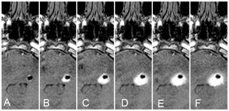
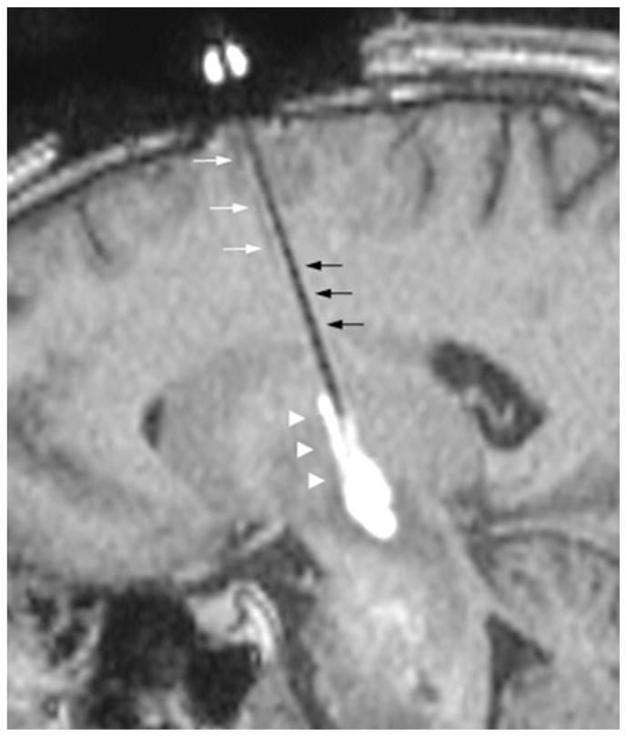
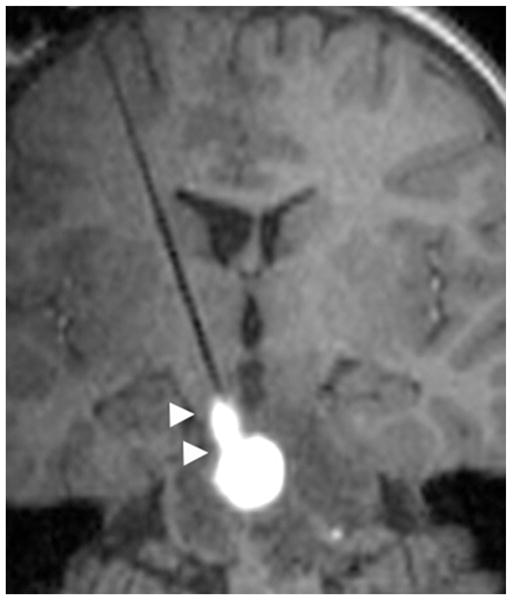
Results: Four patients (2 males, 2 females; mean age at initial infusion 13.0 ± 5.3 years; range 5-17 years) underwent 5 infusions into DIPGs. Brainstem infusions were clearly identified on T1-weighted MR images at 1-mM (1 infusion) and 5-mM (4 infusions) coinfused Gd-DTPA concentrations. While the volume of distribution (Vd) increased progressively with volume of infusion (Vi) (mean volume 2.5 ± 0.9 ml; range 1.1-3.7 ml), final Vd:Vi ratios were significantly reduced with lower Gd-DTPA concentration (Vd:Vi for 1 mM of 1.6 compared with a mean Vd:Vi ratio for 5 mM of 3.3 ± 1.0) (p = 0.04). Similarly, anatomical distribution patterns were affected by preferential flow along parallel axial fiber tracts, into prior infusion cannula tracts and intraparenchymal air pockets, and leak back around the infusion cannula at the highest rate of infusion.
Conclusions: Magnetic resonance imaging of a coinfused Gd-DTPA surrogate tracer provided direct insight into the properties of CED in a clinical application. While clinically relevant Vds can be achieved by convective delivery, specific tissue properties can affect distribution volume and pattern, including Gd-DTPA concentration, preferential flow patterns, and infusion rate. Understanding of these properties of CED can enhance its clinical application. Part of clinical trial no. NCT00880061 ( ClinicalTrials.gov ).
Figures

Comment in
-
Editorial: Convection-enhanced delivery for diffuse intrinsic pontine glioma.J Neurosurg Pediatr. 2014 Mar;13(3):273-4. doi: 10.3171/2013.10.PEDS13421. Epub 2014 Jan 10. J Neurosurg Pediatr. 2014. PMID: 24410123 No abstract available.
-
Response.J Neurosurg Pediatr. 2014 Mar;13(3):274-5. J Neurosurg Pediatr. 2014. PMID: 24724188 No abstract available.
References
-
- Bruce JN, Falavigna A, Johnson JP, Hall JS, Birch BD, Yoon JT, et al. Intracerebral clysis in a rat glioma model. Neurosurgery. 2000;46:683–691. - PubMed
-
- Chen MY, Lonser RR, Morrison PF, Governale LS, Oldfield EH. Variables affecting convection-enhanced delivery to the striatum: a systematic examination of rate of infusion, cannula size, infusate concentration, and tissue–cannula sealing time. J Neurosurg. 1999;90:315–320. - PubMed
-
- Croteau D, Walbridge S, Morrison PF, Butman JA, Vortmeyer AO, Johnson D, et al. Real-time in vivo imaging of the convective distribution of a low-molecular-weight tracer. J Neurosurg. 2005;102:90–97. - PubMed
Publication types
MeSH terms
Substances
Associated data
Grants and funding
LinkOut - more resources
Full Text Sources
Other Literature Sources
Medical
