. 2014 Jun;20(11-12):1632-42.
doi: 10.1089/ten.TEA.2013.0515. Epub 2014 Feb 27.

Biocompatible tissue scaffold compliance promotes salivary gland morphogenesis and differentiation

Affiliations

Biocompatible tissue scaffold compliance promotes salivary gland morphogenesis and differentiation

Sarah B Peters et al. Tissue Eng Part A. 2014 Jun.

Abstract

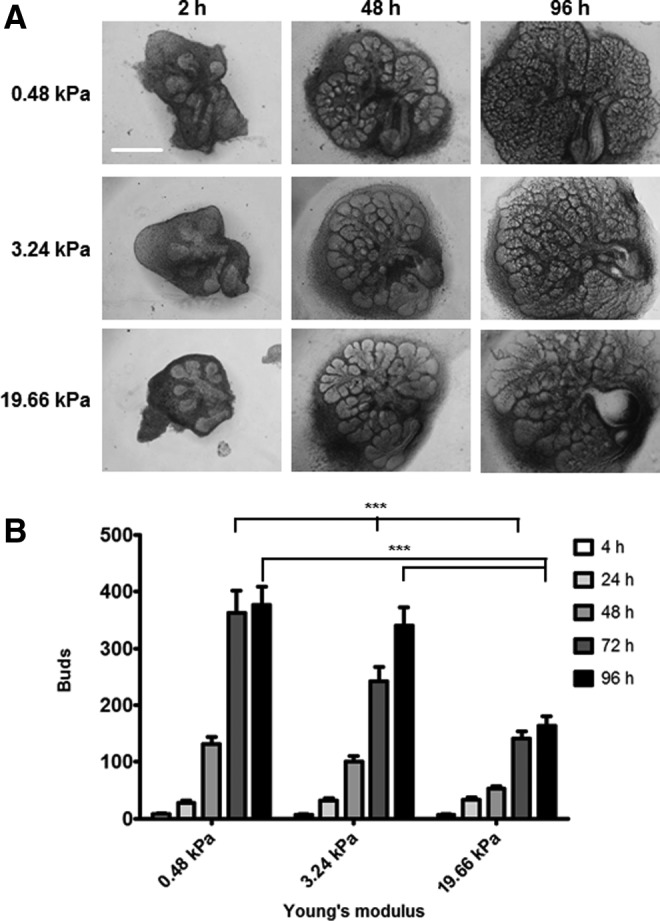
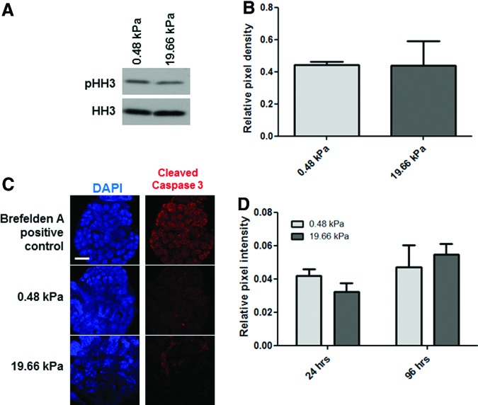
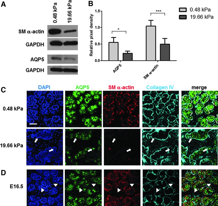
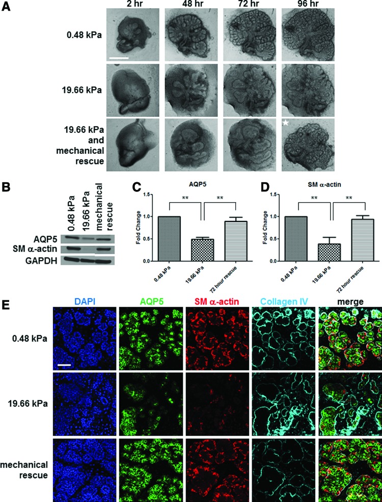
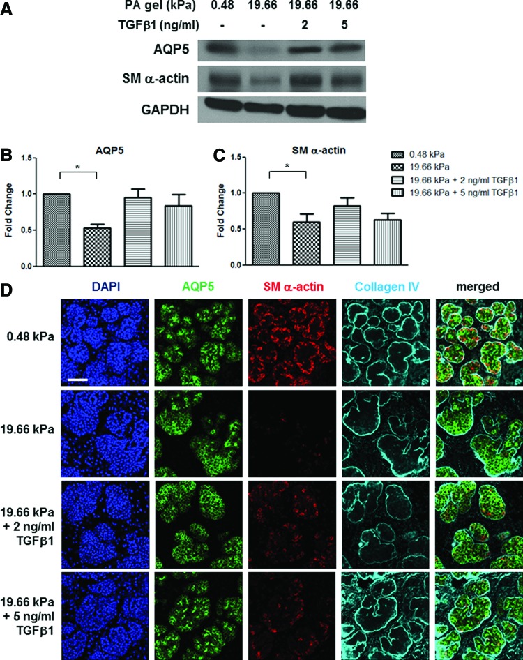
Substrate compliance is reported to alter cell phenotype, but little is known about the effects of compliance on cell development within the context of a complex tissue. In this study, we used 0.48 and 19.66 kPa polyacrylamide gels to test the effects of the substrate modulus on submandibular salivary gland development in culture and found a significant decrease in branching morphogenesis in explants grown on the stiff 19.66 kPa gels relative to those grown on the more physiologically compliant 0.48 kPa gels. While proliferation and apoptosis were not affected by the substrate modulus, tissue architecture and epithelial acinar cell differentiation were profoundly perturbed by aberrant, high stiffness. The glands cultured on 0.48 kPa gels were similar to developing glands in morphology and expression of the differentiation markers smooth muscle alpha-actin (SM α-actin) in developing myoepithelial cells and aquaporin 5 (AQP5) in proacinar cells. At 19.66 kPa, however, tissue morphology and the expression and distribution of SM α-actin and AQP5 were disrupted. Significantly, aberrant gland development at 19.66 kPa could be rescued by both mechanical and chemical stimuli. Transfer of glands from 19.66 to 0.48 kPa gels resulted in substantial recovery of acinar structure and differentiation, and addition of exogenous transforming growth factor beta 1 at 19.66 kPa resulted in a partial rescue of morphology and differentiation within the proacinar buds. These results indicate that environmental compliance is critical for organogenesis, and suggest that both mechanical and chemical stimuli can be exploited to promote organ development in the contexts of tissue engineering and organ regeneration.


Figures

<b>FIG. 1.</b>
FIG. 1.
Branching morphogenesis is promoted by a compliant substrate. (A, B) Branching decreases with stiffness. (A) Representative brightfield images exhibit E13 glands having six to seven buds that were grown on PA gels of three moduli for up to 96 h; scale bar=500 μm. (B) Morphometric analysis to quantify bud numbers was performed for organ explants grown on the three different PA gel moduli at 24-h increments. A two-way ANOVA test with Bonferroni post-tests was applied at each time point (n=8 explants/condition; ***p<0.001). Differences between bars not marked are not statistically relevant. ANOVA, analysis of variance; E13, embryonic day 13; PA, polyacrylamide.
<b>FIG. 2.</b>
FIG. 2.
Decreased branching morphogenesis at high stiffness is not due to differences in proliferation or apoptosis. (A, B) Proliferation. (A) Western detection of phospho-histone H3 (pHH3) in comparison with total histone H3 (HH3) demonstrates no significant difference in the levels of phosphorylation at Ser 10 between glands grown on soft (0.48 kPa) and stiff (19.66 kPa) PA gels for 96 h. (B) Quantification of western analysis: relative pixel density of pHH3 relative to HH3. One-way ANOVA indicates no significant difference between the samples (n=3 experiments, p>0.05). (C, D) Apoptosis. (C) Single confocal images captured from representative organ explants cultured for 96 h and subjected to ICC to detect cleaved caspase 3 and counterstained with DAPI; scale bar=250 μm. Addition of brefelden A to culture media to glands grown atop polycarbonate filters induced apoptosis as a positive control that was used to define cleaved caspase-3-positive nuclei in morphometric analysis of pixel intensity in (D). (D) Quantification of apoptotic nuclei expressed as cleaved caspase-3-positive nuclei relative to total nuclei (DAPI) from stacks of confocal images spanning the thickness of the gland. One-way ANOVA indicates no significant difference in apoptosis between glands cultured on 0.48 kPa and 19.66 kPa gels after 24 or 96 h of culture (n=4 explants/condition, p>0.05). ICC, immunocytochemistry. Color images available online at www.liebertpub.com/tea
<b>FIG. 3.</b>
FIG. 3.
Salivary gland proacinar morphology and epithelial differentiation are physiologically advanced by a compliant substrate and disrupted by aberrant stiffness. (A, B) Expression levels of differentiation markers. (A) Western analysis indicates a decrease in protein levels of both aquaporin 5 (AQP5) and SM α-actin with increasing stiffness. (B) Quantification of western analysis for AQP5 and SM α-actin protein levels, graphed as the relative pixel density of each band relative to the GAPDH control. A two-way ANOVA test with Bonferroni post-tests was applied at each time point (n=6 experiments, *p<0.05 for AQP5 and ***p<0.001 for SM α-actin). (C) Explant gland morphology and differentiated cell arrangement. Representative single confocal images captured from the center of organ explants demonstrate rounded buds, as detected by ICC for the basement membrane protein collagen IV (cyan) in explants grown on the 0.48 kPa gels. Less well-organized and less-consistent morphology is evident in explants cultured on 19.66 kPa gels. The myoepithelial cell protein SM α-actin (red) is expressed in the outer cuboidal cell populations of the rounded buds, and the proacinar/acinar protein AQP5 (green) is expressed in the interior epithelial cells in the glands cultured at low stiffness. In explants cultured at 19.66 kPa, the less-homogeneous bud structures generally show decreases in AQP5 and SM α-actin protein levels. Additionally, apically localized AQP5 can be detected in aberrant acinar structures lacking SM α-actin-positive cells (white arrows). n=10 experiments. (D) In vivo gland morphology and differentiated cell arrangement. Single confocal images captured at the center of E16.5 proacinar buds demonstrate that AQP5 is localized apically by the inner epithelial cells (green), highlighted by arrow heads, as in glands grown on 0.48 kPa PA gels. SM α-actin (red) is expressed in the outer cuboidal cells of the proacinar structures, interior to the basement membrane, as detected by anti-Col IV antibody (cyan). Scale bar=50 μm. E16.5, embryonic day 16.5; SM α-actin, smooth muscle alpha-actin.
<b>FIG. 4.</b>
FIG. 4.
The aberrant development of salivary gland organ explants grown on a stiff substrate can be rescued with a compliant substrate. (A) Mechanical rescue of branching morphogenesis. Brightfield images show representative E13 salivary glands grown at 0.48 and 19.66 kPa for 96 h with disruption of both bud morphology and number that is evident at 19.66 kPa. When transferred to a 0.48 kPa PA gel after 72 h culture on a 19.66 kPa gel, the glands regain a normal bud morphology and number similar to glands cultured on 0.48 kPa PA gels continuously. The white star indicates a transferred gland; scale bar=500 μm. (B–D) Mechanical rescue of epithelial differentiation marker expression levels. (B) Western analysis indicates a decrease in both AQP5 and SM α-actin with increasing stiffness. There is a near-complete rescue of both AQP5 and SM α-actin protein levels when glands are transferred from 19.66 kPa gel to the compliant 0.48 kPa gel. (C, D) Quantification of the western analysis of AQP5 (C) and SM α-actin (D), normalized to GAPDH and graphed as the fold change relative to the 0.48 kPa pixel density. A one-way ANOVA test with Bonferroni post-tests was applied for each protein (n=4 experiments, **p<0.01). (E) Mechanical rescue of gland morphology and differentiated cell arrangement. Representative confocal images of glands transferred from 19.66 to 0.48 kPa demonstrate a robust rescue in bud morphology, as indicated by DAPI (blue) and collagen IV (cyan) localization and redistribution of both AQP5 (green) and SM α-actin (red). n=3 experiments. Scale bar=50 μm. Color images available online at www.liebertpub.com/tea
<b>FIG. 5.</b>
FIG. 5.
The aberrant development of salivary glands grown on high-stiffness substrates is partially rescued with exogenous TGFβ1. (A–C) Chemical rescue of epithelial differentiation marker expression levels. (A) Western analysis indicates a decrease in AQP5 and SM α-actin with increasing stiffness that is partially rescued with exogenous TGFβ1 added to the culture media. (B, C) Quantification of western analysis to detect AQP5 and SM α-actin in response to TGFβ1, normalized to GAPDH and expressed as the fold change relative to the 0.48 kPa value. A one-way ANOVA test with Bonferroni post-tests was applied for each protein (n=5 experiments, *p<0.05, p>0.05 for all other comparisons). TGFβ1 at either 2 or 5 ng/mL concentration did not significantly stimulate total protein levels of AQP5 or SM α-actin. (D) Chemical rescue of gland morphology and bilayered acini. Representative confocal images demonstrate that exogenous TGFβ1 (2 ng/mL) added to glands cultured on the high-stiffness substrate stimulates restoration of acinar morphology and uniformity, as detected by nuclear distribution (DAPI) and collagen IV (cyan) distribution concomitant with increased localization of SM α-actin (red) to the outer periphery of the proacinar buds. Additionally, with 5 ng/mL of exogenous TGFβ1, AQP5 (green) is progressively apically localized in the central cells together with the increased localization of SM α-actin in the outer cells in uniform proacinar structures. n=3 experiments. Scale bar=50 μm. TGFβ1, transforming growth factor beta 1. Color images available online at www.liebertpub.com/tea

Similar articles

Cited by

References

    1. Vacanti C.The history of tissue engineering. J Cell Mol Med 1,569, 2006 - PMC - PubMed
    1. Atala A.Recent developments in tissue engineering and regenerative medicine. Curr Opin Pediatr 18,167, 2006 - PubMed
    1. Atala A., Bauer S.B., Soker S., Yoo J.J., and Retik A.B.Tissue-engineered autologous bladders for patients needing cystoplasty. Lancet 367,1241, 2006 - PubMed
    1. Vrana N.E., Lavalle P., Dokmeci M.R., Dehghani F., Ghaemmaghami A.M., and Khademhosseini A.Engineering functional epithelium for regenerative medicine and in vitro organ models: a review. Tissue Eng Part B Rev 19,529, 2013 - PMC - PubMed
    1. Daley W.P., Peters S.B., and Larsen M.Extracellular matrix dynamics in development and regenerative medicine. J Cell Sci 336,169, 2008 - PubMed

Publication types

MeSH terms

LinkOut - more resources