. 2014 Jan 10;56(1):3.
doi: 10.1186/1751-0147-56-3.

Application of optical coherence tomography enhances reproducibility of arthroscopic evaluation of equine joints

Affiliations

Application of optical coherence tomography enhances reproducibility of arthroscopic evaluation of equine joints

Tytti Niemelä et al. Acta Vet Scand. .

Abstract

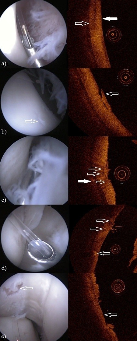
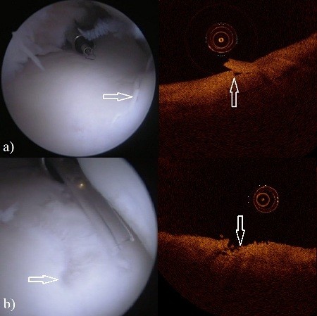
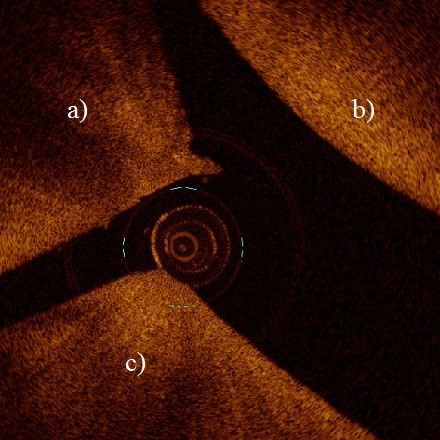
Background: Arthroscopy is widely used in various equine joints for diagnostic and surgical purposes. However, accuracy of defining the extent of cartilage lesions and reproducibility in grading of lesions are not optimal. Therefore, there is a need for new, more quantitative arthroscopic methods. Arthroscopic optical coherence tomography (OCT) imaging is a promising tool introduced for quantitative detection of cartilage degeneration and scoring of the severity of chondral lesions. The aim of this study was to evaluate the inter-investigator agreement and inter-method agreement in grading cartilage lesions by means of conventional arthroscopy and with OCT technique. For this aim, 41 cartilage lesions based on findings in conventional and OCT arthroscopy in 14 equine joints were imaged, blind coded and independently ICRS (International Cartilage Repair Society) scored by three surgeons and one PhD-student.

Results: The intra- and inter-investigator percentages of agreement by means of OCT (68.9% and 43.9%, respectively) were higher than those based on conventional arthroscopic imaging (56.7% and 31.7%, respectively). The intra-investigator Kappa coefficients were 0.709 and 0.565 for OCT and arthroscopy, respectively. Inter-investigator Kappa coefficients were 0.538 and 0.408 for OCT and arthroscopy, respectively.

Conclusions: OCT can enhance reproducibility of arthroscopic evaluation of equine joints.


Figures

Figure 1
Figure 1
The arthroscopy (left) and OCT (right) images of the metacarpo-/metatarsophalangeal joint. The circular object in OCT images is the OCT catheter (diameter = 0.9 mm). a) Dorsal aspect of the lateral sesamoid bone. In the arthroscopic view the tip of the custom made hollow instrument with OCT catheter lie on the examined cartilage surface (ICRS score 0). In the OCT image the articular surface (white arrow) and the interface between cartilage and subchondral bone are visible (open arrow). b) In the arthroscopic view cartilage of the dorsal aspect of the lateral sesamoid bone with lesion (open arrow) is visualized, synovial villi can be seen in the right. The OCT image shows the same lesion (ICRS score 2) with cartilage flap (open arrow) still attached. c) In the arthroscopic view cartilage of the dorsal aspect of the medial sesamoid bone with roughening of the surface is in the left, synovial villi are in the right. The OCT image shows the fibrillation with high definition (ICRS score 2). Beneath the cartilage surface there is cavitation (open arrows) and within the subchondral bone an anomaly (white arrow) can be seen. d) In the view of an arthroscope dorsolateral condyle of third metacarpal bone with cartilage lesion below the OCT catheter can be seen. In the OCT image the lesion (ICRS score 1) and also anomalies beneath the cartilage surface are visible (arrows). e) Severe lesion (ICRS score 4, arrow) of medial sesamoid bone with complete loss of cartilage can be seen in both arthroscope and OCT images.
Figure 2
Figure 2
The arthroscopy (left) and OCT (right) images of the third carpal bone in the intercarpal joint. a) Lesion (arrows) in the arthroscopic image (left) and in the OCT image (right) with ICRS score of 2. b) Lesion (arrows) in the arthroscopic image (left) and in the OCT image (right) with ICRS score of 3.
Figure 3
Figure 3
OCT enables access to the palmar and plantar areas in distal interphalangeal joint which is not achievable with conventional arthroscopy. Image shows the palmar aspect of the distal interphalangeal joint where dorsal aspect of the navicular bone (a) and part of the second (b) and third (c) phalanx are visible.
Figure 4
Figure 4
Mild cartilage fibrillation on the surface of the patella (ICRS score 1) visible only in OCT image (right).

References

    1. Neundorf RH, Lowerison MB, Cruz AM, Thomason JJ, McEven BJ, Hurtig MB. Determination of the prevalence and severity of metacarpophalangeal joint osteoarthritis in Thoroughbred racehorses via quantitative macroscopic evaluation. Am J Vet Res. 2010;56:1284–1293. doi: 10.2460/ajvr.71.11.1284. - DOI - PubMed
    1. Brittberg M, Winalski CS. Evaluation of cartilage injuries and repair. J Bon Joint Surg. 2003;56(Suppl 2):58–69. - PubMed
    1. Verwilghen DR, Enzerink E, Martens A, Franck T, Balligand M, Henrotin Y, Detilleux J, Serteyn D. Relationship between arthroscopic joint evaluation and the levels of Coll2-1, Coll2-1NO2, and myeloperoxidase in the blood and synovial fluid of horses affected with osteochondrosis of the tarsocrural joint. Osteoarthr Cartil. 2011;56:1323–1329. doi: 10.1016/j.joca.2011.08.002. - DOI - PubMed
    1. Spahn G, Klinger HM, Baums M, Pinkepank U, Hofman GO. Reliability in arthroscopic grading of cartilage lesions. Results of a prospective blinded study for evaluation of inter-observer reliability. Arch Orthop Traum Su. 2011;56:377–381. doi: 10.1007/s00402-011-1259-8. - DOI - PubMed
    1. Spahn G, Klinger HM, Hofman GO. How valid is the arthroscopic diagnosis of cartilage lesions? Results of an opinion survey among highly experienced arthroscopic surgeons. Arch Orthop Traum Su. 2009;56:1117–1121. doi: 10.1007/s00402-009-0868-y. - DOI - PMC - PubMed

Publication types