. 2014 Mar 1;88(3):677-85.
doi: 10.1016/j.ijrobp.2013.11.215. Epub 2014 Jan 7.

Acquired tumor cell radiation resistance at the treatment site is mediated through radiation-orchestrated intercellular communication

Affiliations

Acquired tumor cell radiation resistance at the treatment site is mediated through radiation-orchestrated intercellular communication

Natarajan Aravindan et al. Int J Radiat Oncol Biol Phys. .

Abstract

Purpose: Radiation resistance induced in cancer cells that survive after radiation therapy (RT) could be associated with increased radiation protection, limiting the therapeutic benefit of radiation. Herein we investigated the sequential mechanistic molecular orchestration involved in radiation-induced radiation protection in tumor cells.

Results: Radiation, both in the low-dose irradiation (LDIR) range (10, 50, or 100 cGy) or at a higher, challenge dose IR (CDIR), 4 Gy, induced dose-dependent and sustained NFκB-DNA binding activity. However, a robust and consistent increase was seen in CDIR-induced NFκB activity, decreased DNA fragmentation, apoptosis, and cytotoxicity and attenuation of CDIR-inhibited clonal expansion when the cells were primed with LDIR prior to challenge dose. Furthermore, NFκB manipulation studies with small interfering RNA (siRNA) silencing or p50/p65 overexpression unveiled the influence of LDIR-activated NFκB in regulating CDIR-induced DNA fragmentation and apoptosis. LDIR significantly increased the transactivation/translation of the radiation-responsive factors tumor necrosis factor-α (TNF-α), interleukin-1α (IL-1α), cMYC, and SOD2. Coculture experiments exhibit LDIR-influenced radiation protection and increases in cellular expression, secretion, and activation of radiation-responsive molecules in bystander cells. Individual gene-silencing approach with siRNAs coupled with coculture studies showed the influence of LDIR-modulated TNF-α, IL-1α, cMYC, and SOD2 in induced radiation protection in bystander cells. NFκB inhibition/overexpression studies coupled with coculture experiments demonstrated that TNF-α, IL-1α, cMYC, and SOD2 are selectively regulated by LDIR-induced NFκB.

Conclusions: Together, these data strongly suggest that scattered LDIR-induced NFκB-dependent TNF-α, IL-1α, cMYC, and SOD2 mediate radiation protection to the subsequent challenge dose in tumor cells.


Conflict of interest statement

Conflict of interest: none.

Figures

Fig. 1
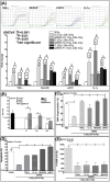
Fig. 1
(A) NFκB DNA-binding activity in cells exposed to mock IR or LDIR and analyzed after 1 hour. Densitometry shows LDIR-induced dose-dependent increase in NFκB activity. (B) Histograms show specificity of DNA-binding activity. (C) Western blot shows levels of phosphorylated IκBα after 1 hour in cells exposed to LDIR. Densitometry shows dose-dependent induction of pIκBα in LDIR-exposed cells. (D) Kinetics of NFκB DNA-binding activity (EMSA) in cells exposed to LDIR at 3 to 24 hours post-IR. Densitometry shows LDIR-induced consistent increases in NFκB activity. (E) cIAP1, cIAP2, survivin, and Bcl2 transactivation (QPCR) in cells exposed to LDIR at 24 hours post-IR. (F) Increased expression levels are shown of IAP1, IAP2, and survivin in cells exposed to LDIR after 24 hours. Densitometry shows induction of IAP1, IAP2, and survivin in cells exposed to LDIR. (G) MTT analysis shows significant increase in survival in cells exposed to 10, 50, or 100 cGy in contrast to mock irradiation. (H) Computed colony counting (Image Quant) shows clonogenic activity of cells either mock irradiated or exposed to LDIR. EMSA = electrophoretic mobility shift assay; IR = ionizing radiation; LDIR = low-dose irradiation; MTT = tetrazolium dye MTT 3-(4,5-dimethylthiazol-2-yl)-2,5-diphenyltetrazolium bromide; QPCR = quantitative realtime plymerase chain reaction.
Fig. 2
Fig. 2
(A) NFκB DNA binding activity in cells exposed to CDIR (4Gy) or primed with LDIR (10, 50, or 100 cGy) followed by CDIR is shown. Densitometry showed increased NFκB DNA binding activity after CDIR. However, LDIR priming robustly increased CDIR-induced NFκB. (B) Kinetics of LDIR priming-associated increase in CDIR-induced NFκB DNA-binding activity after 3 hours through 72 hours post-CDIR are shown. Densitometry shows LDIR-associated robust increase in CDIR-induced NFκB activity remained consistent at least up to 72 hours. (C) Flow cytometry shows complete inhibition of CDIR-induced DNA fragmentation in cells primed with LDIR. (D) Flow cytometry shows the influence of LDIR priming-induced NFκB in inhibiting CDIR-induced DNA fragmentation. NFκB overexpression prevented CDIR-induced DNA fragmentation. Likewise, muting LDIR-induced NFκB brought back CDIR-induced DNA fragmentation consistently up to 48 hours post-CDIR. (E) Flow cytometry of annexin V-FITC staining shows modulation in apoptosis in cells exposed to CDIR with/without LDIR priming or NFκB overexpression and in NFκB-muted LDIR-primed cells exposed to CDIR. (F) Alterations in caspase-3 and -7 activity in cells exposed to CDIR with/without LDIR priming. (G) MTT analysis shows cell survival after CDIR with/without LDIR priming. (H) Clonogenic activity of cells exposed to CDIR with/without LDIR priming is shown. CDIR = challenge-dose irradiation; FITC = fluorescein isothiocyanate; LDIR = low-dose irradiation; MTT = tetrazolium dye MTT 3-(4,5-dimethylthiazol-2-yl)-2,5-diphenyltetrazolium bromide.
Fig. 3
Fig. 3
(A) TNF-α, IL-1α, SOD2, and cMYC transactivation (QPCR) in LDIR-exposed cells at 24 hours post-LDIR. (B) Immunoblots show expression levels of TNF-α, IL-1α, SOD2, and cMYC in cells exposed to LDIR. Densitometry shows significant induction of TNF-α, IL-1α, SOD2, and cMYC in cells after LDIR. (C-G) Coculture experiments show LDIR-induced translation of TNF-α, IL-1α, SOD2, and cMYC in bystander cells and associated radiation protection. (C) SOD2 activity in cells exposed to CDIR with or without LDIR priming is shown. Coculturing CDIR and LDIR-exposed cells significantly induced CDIR-regulated SOD2 activity in bystander cells. (D) ELISA shows secreted TNF-α in cells exposed to CDIR with/without coculturing with LDIR-treated cells. Coculturing significantly increased TNF-α secretion in bystander cells. (E) Immunoblots show expression levels of TNF-α, IL-1α, SOD2, pIκBα, and cMYC in cells exposed to CDIR with/without coculturing with LDIR-treated cells. (F) Cell viability in cells exposed to CDIR with/without coculturing with LDIR-treated cells is shown. (G) Cell survival is shown in cells exposed to CDIR with/without coculturing with LDIR-treated cells. (H) LDIR-induced TNF-α, IL-1α, and RelA transactivation (QPCR) in nontargeted bystander cells across tumor models, including neuroblastoma (SH-SY5Y), breast (MCF-7, MDA-MB-435, MDA-MB-468), bladder (TCC-SUP, J82), colon/gastric (Colo-205, AGS), prostate (DU-145), and lung (A549) cancer cell lines. LDIR = low-dose irradiation; QPCR = quantitative realtime plymerase chain reaction.
Fig. 4
Fig. 4
(A) LDIR-induced NFκB mediates TNF-α, SOD2, cMYC, and IL-1α transactivation in bystander cells. Cells cocultured with NFκB-silenced, LDIR-exposed cells were then exposed to CDIR and analyzed 24 hours post-LDIR. Silencing LDIR-induced NFκB inhibited LDIR priming-associated increases in TNF-α, SOD2, cMYC, and IL-1α in bystander cells. (B) LDIR-induced NFκB mediates increased SOD2 activity in cocultured CDIR-exposed bystander cells. (C-E) LDIR-translated TNF-α, SOD2, cMYC, and IL-1α mediated radiation protection in bystander cells. Gene-specific knockout cells cocultured with/without LDIR were then exposed to CDIR and analyzed for (C) DNA fragmentation, (D) apoptosis, and (E) cell survival. Knocking out radiation-responsive TNF-α, SOD2, cMYC, and IL-1α in bystander cells profoundly reverted LDIR priming-inhibited DNA fragmentation and apoptosis and inhibited LDIR priming-induced cell survival. CDIR = challenge-dose irradiation; LDIR = low-dose irradiation.

References

    1. American Cancer Society. Cancer facts and figures 2012. Atlanta: American Cancer Society; 2012.
    1. Berkey FJ. Managing the adverse effects of radiation therapy. Am Fam Physician. 2010;82:381–388. - PubMed
    1. Russell J, Wheldon TE, Stanton P. A radioresistant variant derived from a human neuroblastoma cell line is less prone to radiation-induced apoptosis. Cancer Res. 1995;55:4915–4921. - PubMed
    1. Stecca C, Gerber GB. Adaptive response to DNA-damaging agents: A review of potential mechanisms. Biochem Pharmacol. 1998;55:941–951. - PubMed
    1. Madhusoodhanan R, Natarajan M, Veeraraghavan J, et al. NFκB activity and transcriptional responses in human breast adenocarcinoma cells after single and fractionated irradiation. Cancer Biol Ther. 2009;8:765–773. - PubMed

Publication types

MeSH terms