. 2014 Mar;35(9):2664-79.
doi: 10.1016/j.biomaterials.2013.11.078. Epub 2014 Jan 8.

Three-dimensional scaffolds of acellular human and porcine lungs for high throughput studies of lung disease and regeneration

Affiliations

Three-dimensional scaffolds of acellular human and porcine lungs for high throughput studies of lung disease and regeneration

Darcy E Wagner et al. Biomaterials. 2014 Mar.

Abstract

Acellular scaffolds from complex whole organs such as lung are being increasingly studied for ex vivo organ generation and for in vitro studies of cell-extracellular matrix interactions. We have established effective methods for efficient de and recellularization of large animal and human lungs including techniques which allow multiple small segments (∼ 1-3 cm(3)) to be excised that retain 3-dimensional lung structure. Coupled with the use of a synthetic pleural coating, cells can be selectively physiologically inoculated via preserved vascular and airway conduits. Inoculated segments can be further sliced for high throughput studies. Further, we demonstrate thermography as a powerful noninvasive technique for monitoring perfusion decellularization and for evaluating preservation of vascular and airway networks following human and porcine lung decellularization. Collectively, these techniques are a significant step forward as they allow high throughput in vitro studies from a single lung or lobe in a more biologically relevant, three-dimensional acellular scaffold.

Keywords: Acellular matrix; Endothelial cell; Epithelial cell; Extracellular matrix (ECM); Human lung fibroblast; Mesenchymal stem cell.


Figures

Figure 1
Figure 1. Strategies for development of three-dimensional scaffolds of acellular human and porcine lungs for high throughput analysis
Following whole organ or lobar perfusion decellularization, three-dimensional segments of acellular lung can be excised which retain their native airway and vascular architecture. Small airways and vessels are easily identifiable and can be cannulated to allow for vascular or airway cell inoculations when a calcium alginate coating is used to seal the segment as an artificial pleura. These segments can then be further sliced for high throughput analysis and cells are shown to be retained in the vasculature following vascular inoculation (CBFs) or in the airspaces following airway inoculation (HBEs). Cells can be seen to be maintained in their inoculated compartments, indicating that the integrity of these individual networks is largely maintained following decellularization.
Figure 2
Figure 2. Gross evaluation of cadaveric human lobar and porcine lung decellularization methods
A) Representative gross images at different steps throughout CP decellularization of human lobes (1L/min (CP1), 2L/min (CP2), 3L/min (CP3)). By gross inspection, all scaffolds appeared decellularized at the conclusion of the protocol, as assessed by loss of pink pigmentation following terminal sterilization with peracetic acid (PAA), irrespective of flow rate utilized. B) Bronchoscopy still images of acellular human and porcine lung show maintenance of large airways and removal of cellular material by gross inspection following decellularization. Images represent time lapse captures at major branching points of the proximal and distal bronchi. * indicates the path of the bronchoscope. (See Supplementary Movie 1 for video) C) Thermographic imaging is a powerful tool for examining perfusion pathways in real time during tissue decellularization and for evaluating preservation of airway and vascular systems in acellular scaffolds. Representative still images from thermographic analysis demonstrate maintenance of airway and vasculature using infrared imaging following human lobar decellularization at 2L/min instillation (CP2).Online supplement contains comparable still image panels following decellularization at 1L/min and 3L/min in acellular human lobes (See Supplementary Figs. 2C and 2D for still images of airway instillation at 1L/min or 2L/min into the bronchi of acellular human lobes, respectively). Human lobes were chilled at 4°C in PBS after completion of the decellularization protocol. DI water was warmed to 37°C to create a thermal gradient (temperature scale bar shown) for detection by the FLIR camera. Qualitatively, the branching architecture of the acellular scaffold is largely maintained and proximal followed by distal regions can be seen receiving the warmed DI water solutions as evidenced by changes in surface temperature (indicated by light blue to red color changes). (See Supplementary Movie 2, available online, showing principles of thermography and its use for monitoring perfusion instillation of detergents throughout decellularization of a whole porcine lung at 2L/min.).
Figure 3
Figure 3. Cadaveric human lung lobes lack visible nuclear material following lobar perfusion decellularization and retain major ECM components by histologic and immunohistochemical assessment
A) Representative histologic photomicrographs demonstrate maintenance of histologic architecture and collagen content (pink in H&E and EVG staining, blue in trichrome staining) with all three perfusion flow rates (CP1, CP2, and CP3) in human lung lobes. No viable cells or nuclei (dark purple in H&E and trichrome staining) are evident in any CP method. Qualitatively, the 2L/min and 3L/min flow rates result in increased qualitative loss of elastin (black staining in EVG panels) following decellularization. Decreased cytoplasmic staining (i.e. light red/pink in trichrome) is evident at 2L/min and 3L/min flow rates as qualitatively compared to 1L/min, indicating more complete removal of cellular debris. a = airways, bv = blood vessels. Arrows highlight individual blood vessels. Original magnifications 100X. * Inserts at 200X. (See Supplementary Figs. 3A and 3B for representative histology of manual and VP decellularization of human lobes. See Supplementary Fig. 3C for representative H&E of all methods for porcine lungs.) B) Representative immunofluorescent photomicrographs demonstrate maintenance of ECM proteins and architecture following decellularization using CP methods. As consistent with histologic evaluation, CP2 and CP3 qualitatively results in optimal opening of distal airspaces and blood vessels as compared to CP1. C) Representative immunofluorescent photomicrographs demonstrating retention of smooth muscle actin and smooth muscle myosin in all CP methods. Nuclear DAPI staining is depicted in blue, and the stain(s) of interest are depicted in green in each respective panel. Lam = laminin, Col-1 = type I collagen, Col-4 = type 4 collagen, East = elastin, Fib = fibronectin. SMA = smooth muscle actin, SMM = smooth. Original magnifications 200x.
Figure 4
Figure 4. Use of perfusion pumps and physical agitation during cadaveric human lung lobar decellularization enhances removal of cellular material and preserves architecture
(a) Electron micrographs of human tissue following manual instillation of decellularization reagents confirm that these lungs were heterogeneously decellularized and that significant amounts of cellular debris remain in interstitial spaces. (b) Electron micrographs of human tissue following variable flow rate perfusion instillation of decellularization reagents demonstrates that the use of a perfusion pump for instilling fluids enhances the removal of cell debris and more readily clears debris from small capillaries (labeled “ca”). (c) Electron micrographs of human tissue following constant flow rate perfusion instillation with physical agitation during incubation steps demonstrates more homogeneous clearing of interstitial spaces and capillaries. Lungs decellularized using this method, irrespective of flow rate, appeared to remove intact cellular bodies and retain characteristic normal alveolar septa. Collagen and elastin are indicated with black arrows. Scale bars for EM images are labeled on each inset.
Figure 5
Figure 5. Mass spectrometric assessment of residual proteins following decellularization of human lungs demonstrates that CP1, CP2, and CP3 protocols enhance removal of residual cellular associated proteins as compared to manual instillation of decellularization reagents
A) Positively identified proteins in decellularized human lungs (i.e. those proteins which exceeded the FDR cutoff for identification) from each method of decellularization were assigned to groups according to subcellular location (cytoskeletal, cytosolic, ECM, membrane-associated, nuclear, and secreted). Heatmaps were generated using the log normal transformation of total spectral counts for all positively identified proteins and grouped by category (See Supplementary Fig. 4A for heatmaps of individual acellular human samples from each lobe). Total spectral counts and statistical analyses for all identified proteins by mass spectrometry are included in Supplementary Tables 2–7. The key to human protein identification is in Supplementary Table 8. (Methods of decellularization are described in Table 1; CP1 (n=3 lobes), CP2 (n=4 lobes), and CP3 (n=4 lobes)) B) Three distinct samples were removed from different regions of each lobe decellularized with CP methods and compared to all other individual samples by Spearman’s correlation. Intra-lobe correlation was assessed between individual samples from the same lobe and individual samples derived from single lobes were found to be significantly similar. Samples originated from three different patients for these comparisons. Agglomerative hierarchical clustering of Spearman’s correlation coefficients using the complete linkage method revealed that lung origin (i.e. individual patient) resulted in significant clustering as compared to the flow rate of instillation during decellularization. Therefore, the range of residual proteins following decellularization is specific to each lung. (See Supplementary Fig. 4B for correlation coefficients and Supplementary Fig. 4C for p-values of all comparisons).
Figure 6
Figure 6. Excised segments of acellular human lung can be selectively seeded through airways or vasculature with the use of a calcium alginate coating
A) Small airways and vessels can be identified in acellular segments, cannulated and secured with surgical clips for controlled cellular inoculation. Segments can be coated with a 2.5% sodium alginate solution and ionically crosslinked with a 3% calcium chloride solution to create a hydrogel coating (See Supplementary Fig. 5A). Calcium alginate does not permit cellular adhesion of HLFs and promotes retention of cells in the three-dimensional segment (See Supplementary Figs. 5B and 5C). B) Endothelial progenitor cells (CBF12) inoculated through the vasculature of a calcium alginate coated segment excised from an acellular scaffold decellularized at 2L/min are retained in blood vessels after 24 hours of culture. Human bronchial epithelial cells inoculated through a small airway are retained in the airspaces after 24 hours of culture. Cells were seeded in three-dimensional segments, allowed to adhere overnight, and subsequently sliced and cultured in low attachment tissue culture plates for use in high throughput studies.
Figure 7
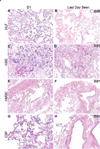
Figure 7. HLF, HBE, CBF, and hMSC cells engraft and remain viable in acellular human lungs
A) HLF, HBE, and hMSC cells inoculated through small airways in acellular human segments and sliced into thin (∼1mm) sections of tissue can be cultured for at least 28 days for high throughput assessments of different culture conditions. Representative H and E photomicrographs depict characteristic recellularization patterns one day after inoculation as well as the last day viable cells could be observed. Subpanels A-H are representative images for each cell type. Engrafted cells in acellular human lungs predominantly acquired characteristic adherent, flattened phenotypes. B) High power images of the last time points observed in acellular human lung slices for each cell type. Original magnification 100x (low power images) and 400x (high power images)

References

    1. Petersen TH, Calle EA, Zhao L, Lee EJ, Gui L, Raredon MB, et al. Tissue-engineered lungs for in vivo implantation. Science. 2010;329:538–541. - PMC - PubMed
    1. Price AP, England KA, Matson AM, Blazar BR, Panoskaltsis-Mortari A. Development of a decellularized lung bioreactor system for bioengineering the lung: the matrix reloaded. Tissue Eng Part A. 2010;16:2581–2591. - PMC - PubMed
    1. Ott HC, Clippinger B, Conrad C, Schuetz C, Pomerantseva I, Ikonomou L, et al. Regeneration and orthotopic transplantation of a bioartificial lung. Nat Med. 2010;16:927–933. - PubMed
    1. Cortiella J, Niles J, Cantu A, Brettler A, Pham A, Vargas G, et al. Influence of acellular natural lung matrix on murine embryonic stem cell differentiation and tissue formation. Tissue Eng Part A. 2010;16:2565–2580. - PubMed
    1. Daly AB, Wallis JM, Borg ZD, Bonvillain RW, Deng B, Ballif BA, et al. Initial binding and recellularization of decellularized mouse lung scaffolds with bone marrow-derived mesenchymal stromal cells. Tissue Eng Part A. 2012;18:1–16. - PMC - PubMed

Publication types

MeSH terms