Clinical Trial
. 2014 Jan 7;19(1):96-108.
doi: 10.1016/j.cmet.2013.12.003.

β-Aminoisobutyric acid induces browning of white fat and hepatic β-oxidation and is inversely correlated with cardiometabolic risk factors

Affiliations
Clinical Trial

β-Aminoisobutyric acid induces browning of white fat and hepatic β-oxidation and is inversely correlated with cardiometabolic risk factors

Lee D Roberts et al. Cell Metab. .

Abstract

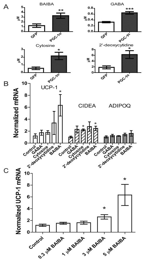
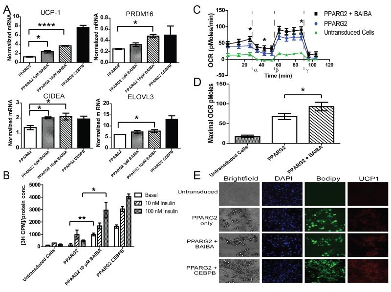
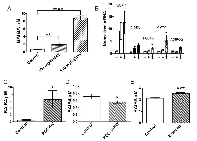
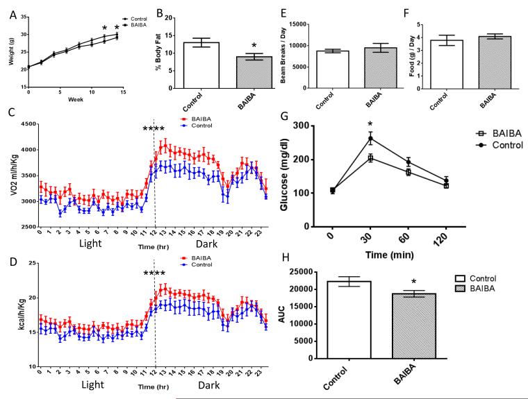
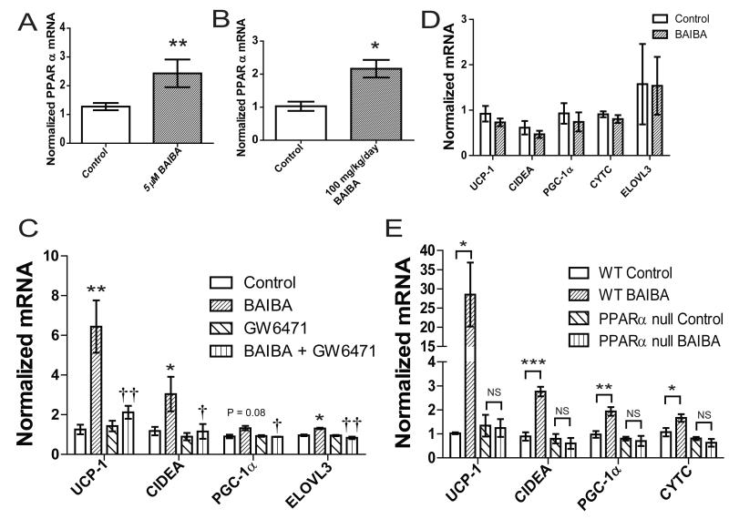
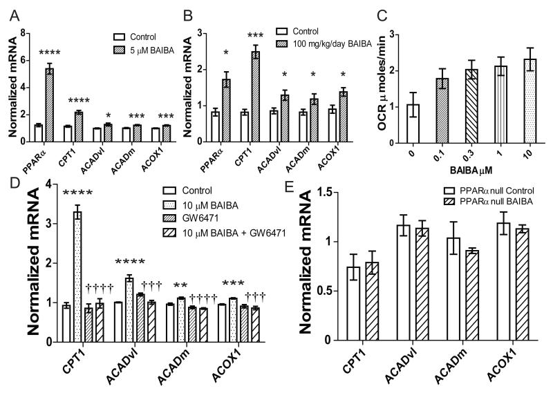
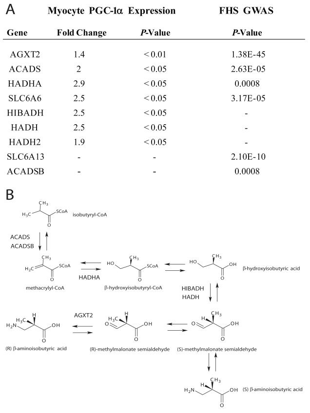
The transcriptional coactivator peroxisome proliferator-activated receptor-gamma coactivator-1α (PGC-1α) regulates metabolic genes in skeletal muscle and contributes to the response of muscle to exercise. Muscle PGC-1α transgenic expression and exercise both increase the expression of thermogenic genes within white adipose. How the PGC-1α-mediated response to exercise in muscle conveys signals to other tissues remains incompletely defined. We employed a metabolomic approach to examine metabolites secreted from myocytes with forced expression of PGC-1α, and identified β-aminoisobutyric acid (BAIBA) as a small molecule myokine. BAIBA increases the expression of brown adipocyte-specific genes in white adipocytes and β-oxidation in hepatocytes both in vitro and in vivo through a PPARα-mediated mechanism, induces a brown adipose-like phenotype in human pluripotent stem cells, and improves glucose homeostasis in mice. In humans, plasma BAIBA concentrations are increased with exercise and inversely associated with metabolic risk factors. BAIBA may thus contribute to exercise-induced protection from metabolic diseases.


Figures

Figure 1
Figure 1. Metabolites accumulate in the media of myocytes as a result of forced PGC-1α expression and stimulate expression of brown adipocyte-specific genes in adipocytes
A) Myocytes were transduced with an adenoviral vector expressing either PGC-1α (n=6) or GFP (n=6). After 24 hours of exposure to these cells, media was analyzed using an LC-MS based metabolite profiling method measuring 100 small molecules (see Methods). B) BAIBA (5 μM) induces expression of brown adipocyte-specific genes in primary adipocytes differentiated from the stromal vascular fraction isolated from inguinal WAT over 6 days. Additional metabolites tested at physiologically relevant doses included GABA (3 μM), cytosine (1 μM), and 2-deoxycytidine (15 μM). While BAIBA significantly increased the expression of the brown adipocyte-specific genes UCP-1 and CIDEA, it did not alter the expression of the white adipocyte gene adiponectin (ADIPOQ). Cumulative data from a total of 5 independent observations are shown. C) BAIBA concentrations in the low micromolar range significantly and dose-dependently increased the expression of the brown adipocyte-specific gene UCP-1. *, P ≤ 0.05, **, P ≤ 0.01, ***, P ≤ 0.001. Data are represented as Mean ± SEM.
Figure 2
Figure 2. BAIBA treatment of BJ RiPS human iPSCs induces brown adipocyte-specific gene expression and function
A) BAIBA significantly and dose dependently increased the expression of brown-adipocyte specific genes in human induced pluripotent stem cell (IPSC) derived mature adipocytes. B) Glucose uptake in human IPSC derived adipocytes was assessed by the transport of [3H]-2-deoxy-D-glucose at basal level and during insulin stimulation (10 nM and 100 nM). BAIBA increased both basal and insulin stimulated glucose uptake (data from 3 independent observations are shown). C) Comparison of the oxygen consumption rate (OCR) of human IPSC-derived white adipocytes with and without BAIBA treatment; Untransduced cells differentiated with adipogenic media (green line), PPARG2 programmed cells (blue line), and PPARG2 programmed cells treated with BAIBA (black line). The OCR was measured over time with the addition of oligomycin (α), an ATPase inhibitor, carbonyl cyanide m-chlorophenyl hydrazone (CCCP), a proton gradient uncoupler (β) allowing determination of the maximum OCR, and antimycin (γ). D) BAIBA significantly increased the maximal OCR of PPARG2 transduced adipocytes. (Data from n = 10 independent observations are shown). E) Images of untransduced cells, PPARG2 programmed white adipocytes, PPARG2 programmed white adipocytes treated with BAIBA, and PPARG2-CEBPB programmed brown adipocytes. Shown from left to right: brightfield images illustrating the morphology of the cells; 4′,6-diamidino-2-phenylindole (DAPI) fluorescent nuclear staining (blue); fluorescent staining with the neutral lipid dye BODIPY (green); fluorescent images of immunostaining with antibodies against the marker protein UCP-1 (red) (100× magnification). *, P ≤ 0.05, **, P ≤ 0.01, ***, P ≤ 0.001,****, P < 0.0001. Data are represented as Mean ± SEM.
Figure 3
Figure 3. BAIBA induces expression of brown adipocyte-specific genes in WAT in vivo and muscle specific PGC-1α expression and exercise significantly increase plasma BAIBA levels
A) The plasma concentration of BAIBA in mice given 100 mg/kg/day (n = 5) or 170 mg/kg/day (n = 5) of the metabolite in their drinking water significantly increased over 14 days as compared to age matched control mice (n = 5). B) Expression of brown adipocyte-specific genes in inguinal WAT from control mice (-) (n = 5), mice treated with 100 mg/kg/day BAIBA for 14 days (+) (n = 5), or mice treated with 170 mg/kg/day BAIBA for 14 days (++) (n=5). C) Plasma from muscle specific PGC-1α transgenic mice (n = 5) was analyzed using an LC-MS metabolite profiling platform and compared to plasma from age matched control mice (n = 5). D) Plasma from PGC-1α knockout mice (n = 9) was analyzed using LC-MS and compared to plasma from age matched control mice (n = 8) E) Mice were subjected to a 3 week free wheel running exercise regimen (n = 6) or housed as sedentary controls (n=6), and plasma BAIBA levels were assessed by LC-MS. *, P ≤ 0.05, **, P ≤ 0.01, ***, P ≤ 0.001, ****, P < 0.0001. Data are represented as Mean ± SEM.
Figure 4
Figure 4. BAIBA decreases weight gain and improves glucose tolerance in mice
A) The weights of mice given 100 mg/kg/day BAIBA (n = 8) in their drinking water compared to untreated controls (n = 8). B) The percentage body fat of 100 mg/kg/day BAIBA treated mice (n = 8) compared to untreated controls (n = 8). C) Diurnal oxygen consumption of control mice (n = 8) and BAIBA (100 mg/kg/day) treated mice (n = 8). D). Diurnal energy expenditure of control mice (n = 8) and BAIBA (100 mg/kg/day) treated mice (n = 8). E) Activity of control mice (n = 8) and mice treated with 100 mg/kg/day BAIBA .F) Food consumption of control mice and mice treated with 100 mg/kg/day BAIBA. G) Mice treated with 100 mg/kg/day BAIBA for 14 weeks showed significantly improved glucose tolerance as determined by an IPGTT. H) The area under the curve of an IPGTT comparing BAIBA treated mice to untreated controls (Control, n = 8, BAIBA n = 8). *, P < 0.05. Data are represented as Mean ± SEM.
Figure 5
Figure 5. PPARα functions downstream of BAIBA
A) BAIBA (5 μM) induces expression of PPARα in primary adipocytes differentiated from the stromal vascular fraction isolated from inguinal WAT over 6 days. Cumulative data from a total of 6 independent observations are shown. B) Expression of PPARα in inguinal WAT from control mice (n = 5) and mice treated with 100 mg/kg/day BAIBA for 14 days (n = 5). C) Primary adipocytes differentiated from the stromal vascular fraction treated with BAIBA (5 μM) and/or GW6471 for 6 days. The graph shows qPCR of indicated genes. †< 0.05,†† < P0.01 compared to BAIBA treatment. D) BAIBA (5 μM) failed to induce expression of brown adipocyte-specific genes in primary adipocytes differentiated from the stromal vascular fraction isolated from inguinal WAT of PPARα null mice. Cumulative data from a total of 6 independent observations are shown. E) Expression of brown adipocyte-specific genes in inguinal WAT from wild type (WT) control mice (n = 5), WT mice treated with 100 mg/kg/day BAIBA for 14 days (n = 5), PPARα null control mice (n=5) and PPARα null mice treated with 100 mg/kg/day BAIBA for 14 days (n=5). *, P < 0.05, **, P ≤ 0.01 compared to control. Data are represented as Mean ± SEM.
Figure 6
Figure 6. BAIBA increases hepatic β-oxidation through PPARα
A) BAIBA (5 μM) induces expression of fatty acid β-oxidation genes in hepatocytes treated for 6 days (Control = 6, BAIBA 5 μM = 6). B) BAIBA dose-dependently induces expression of hepatic fatty acid β-oxidation genes in vivo. Expression of fatty acid β-oxidation genes in the liver of control mice (n = 5) and mice treated with 100 mg/kg/day BAIBA for 14 days (n = 5). C) BAIBA dose dependently increases the respiration rate of hepatocytes. Hepatocytes were treated with a range of BAIBA concentrations (0, 0.1, 0.3, 1, 3, 10 μM) for 6 days. Maximal oxygen consumption rate (OCR) was induced using FCCP. D) qPCR of key β- oxidation genes in hepatocytes treated with BAIBA and/or PPARα antagonist GW6471 for 6 days. Cumulative data from a total of 5 independent observations are shown. †††, P ≤ 0.001, ††††, P < 0.0001 compared to BAIBA treatment. E) Expression of β-oxidation genes in liver from PPARα null control mice (n = 5) and PPARα null mice treated with 100 mg/kg/day BAIBA for 14 days (n = 5). *, P < 0.05, **, P ≤ 0.01, ***, P ≤ 0.001, ****, P < 0.0001 compared to control. All data are represented as Mean ± SEM.
Figure 7
Figure 7. Integration of human genetic and transcriptional data highlights a role for PGC-1α in BAIBA generation
A) A table of the transcriptional changes in genes associated with the BAIBA biosynthesis pathway in primary myocytes expressing PGC-1α as assessed by expression arrays (left panel). Right panel includes genes in the BAIBA biosynthesis pathway and the significance of their relationship to BAIBA plasma concentrations in the Framingham Heart Study (FHS). B) The BAIBA biosynthesis pathway annotated with the genes increased by forced PGC-1α expression in primary myocytes or identified by the GWAS study.

Comment in

References

    1. Ahfeldt T, Schinzel RT, Lee YK, Hendrickson D, Kaplan A, Lum DH, Camahort R, Xia F, Shay J, Rhee EP, et al. Programming human pluripotent stem cells into white and brown adipocytes. Nat Cell Biol. 2012;14:209–219. - PMC - PubMed
    1. al Yacoub N, Romanowska M, Haritonova N, Foerster J. Optimized production and concentration of lentiviral vectors containing large inserts. J Gene Med. 2007;9:579–584. - PubMed
    1. Aoi W, Naito Y, Hang LP, Uchiyama K, Akagiri S, Mizushima K, Yoshikawa T. Regular exercise prevents high-sucrose diet-induced fatty liver via improvement of hepatic lipid metabolism. Biochem Biophys Res Commun. 2011;413:330–335. - PubMed
    1. Bassel-Duby R, Olson EN. Signaling pathways in skeletal muscle remodeling. Annu Rev Biochem. 2006;75:19–37. - PubMed
    1. Begriche K, Massart J, Abbey-Toby A, Igoudjil A, Letteron P, Fromenty B. Beta-aminoisobutyric acid prevents diet-induced obesity in mice with partial leptin deficiency. Obesity (Silver Spring) 2008;16:2053–2067. - PubMed

Publication types

MeSH terms

Substances