. 2014 Feb 6;53(3):433-43.
doi: 10.1016/j.molcel.2013.12.009. Epub 2014 Jan 9.

The intrinsically disordered Sem1 protein functions as a molecular tether during proteasome lid biogenesis

Affiliations

The intrinsically disordered Sem1 protein functions as a molecular tether during proteasome lid biogenesis

Robert J Tomko Jr et al. Mol Cell. .

Abstract

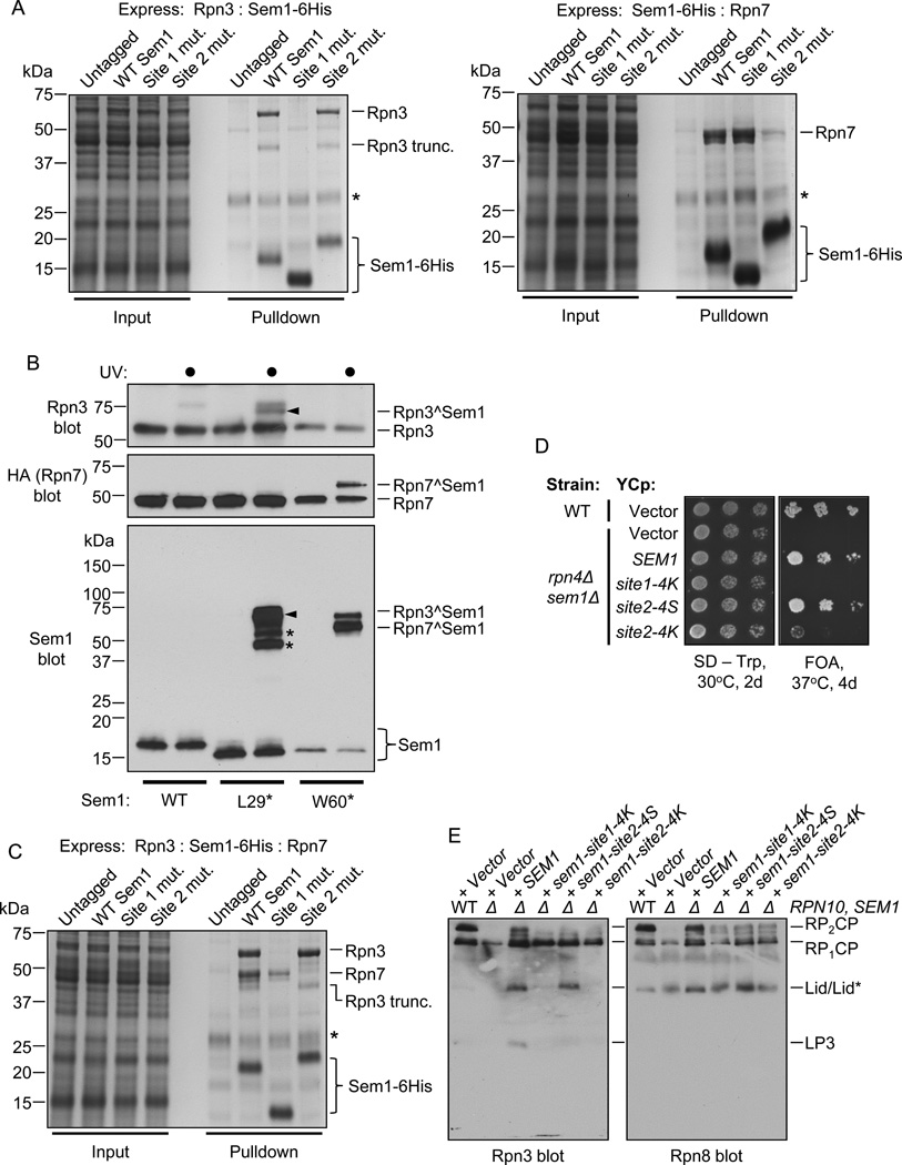
The intrinsically disordered yeast protein Sem1 (DSS1 in mammals) participates in multiple protein complexes, including the proteasome, but its role(s) within these complexes is uncertain. We report that Sem1 enforces the ordered incorporation of subunits Rpn3 and Rpn7 into the assembling proteasome lid. Sem1 uses conserved acidic segments separated by a flexible linker to grasp Rpn3 and Rpn7. The same segments are used for protein binding in other complexes, but in the proteasome lid they are uniquely deployed for recognizing separate polypeptides. We engineered TEV protease-cleavage sites into Sem1 to show that the tethering function of Sem1 is important for the biogenesis and integrity of the Rpn3-Sem1-Rpn7 ternary complex but becomes dispensable once the ternary complex incorporates into larger lid precursors. Thus, although Sem1 is a stoichiometric component of the mature proteasome, it has a distinct, chaperone-like function specific to early stages of proteasome assembly.


Figures

Figure 1
Figure 1. LP3 and Module 1 are competent for assembly
(A) Sequence alignment of Sem1 proteins from the indicated species. Conserved acidic sites 1 and 2 and the poorly conserved linker are indicated. (B) LP3 and Module 1 form a complex that is indistinguishable from LP2. Purified recombinant LP3 and Module 1 were incubated alone or together for 20 min at 30°C before separation by native PAGE and immunoblotting against the indicated lid subunits or their epitope tags. (C) HA-Rpn7 does not purify stoichiometrically with Module 1 (Rpn5/6/8/9/11) in the absence of Rpn3. Module 1 subunits were coexpressed with the indicated proteins. Complexes were purified via MBP-Rpn6, and then subjected to Sephacryl S-200 chromatography. Normalized gel filtration traces are shown. Module 1 subunits eluted identically at approx. 38 mL in all cases. (D) HA-Rpn7 does not purify stoichiometrically with Module 1 subunits when coexpressed without or with Sem1. SDS-PAGE analysis of fractions from the elution peaks shown in (C) at approx. 38 mL. HA-Rpn7 was weakly detectable by immunoblot in these fractions (not shown). (E) HA-Rpn7 copurifies stoichiometrically with Module 1 subunits when Rpn3 and Sem1 are also present. All lid subunits except Rpn12 were coexpressed in E. coli, and complexes were purified via the MBP tag on Rpn6, followed by Superose 6 chromatography. An aliquot of the largest eluted species was resolved on an SDS-PAGE gel and stained with Coomassie Brilliant Blue. No fractions containing a subset of Rpn3, Rpn7, and Sem1 together with Module 1 subunits were recovered (not shown), supporting the idea that they enter the assembling lid together. See also Figure S1.
Figure 2
Figure 2. Sem1 binds both Rpn3 and Rpn7 and is essential for efficient lid assembly in vivo
(A) Dimeric interaction analysis of LP3 subunits. Dots indicate a subunit was coexpressed; 6His-tagged subunits are marked by filled dots. Rpn3 was largely insoluble unless coexpressed with Sem1. Immunoblotting of the same samples with antibodies against Sem1 is shown at the bottom. (B) Rpn3, but not Rpn5 or Rpn8, is absent from a lid-like particle in sem1Δ yeast. Native PAGE-immunoblot of extracts from the indicated strains. Lid* indicates a species that migrates similarly to the fully assembled lid, but is devoid of Rpn3. (C) Rpn7 is absent from lid*. Immunoblots of the indicated strains as in (B), but each strain contained a chromosomal RPN7-6xGly-3xFLAG allele. See also Figures S1 and S2.
Figure 3
Figure 3. Two evolutionarily conserved sites in Sem1 are important for binding Rpn3 and Rpn7
(A) The indicated Sem1 proteins were coexpressed with Rpn3 (left panel) or Rpn7 (right panel). Sem1 and associated proteins were then isolated via TALON affinity purification. Mutation of residues in Sem1 caused shifts in migration upon SDS-PAGE. Asterisks indicate metal-binding bacterial proteins; Site 1 mut.: C-terminally 6His-tagged Sem1 in which residues 30–37 were mutated to alanine; Site 2 mut.: C-terminally 6His-tagged Sem1 in which W60 and W64 were mutated to alanine and threonine, respectively. (B) Site-specific photocrosslinking of Sem1 site 1 and site 2 to Rpn3 and Rpn7, respectively. Sem1-Gly-6His containing p-benzoylphenylalanine at the indicated positions and associated proteins were purified on a TALON resin, followed by UV irradiation (indicated by a black dot) to induce crosslinking. The Sem1^Rpn3 crosslink is indicated by an arrowhead. Sem1-L29* also appeared to crosslink to Rpn3 truncations (indicated by asterisks in the Sem1 blot). (C) Both site 1 and site 2 must be intact for LP3 formation. Rpn3, Rpn7, and the indicated forms of Sem1 were coexpressed and purified as in (A). Asterisk, bacterial metal-binding protein. (D) The indicated yeast strains were transformed with empty vector or low-copy plasmids encoding the indicated SEM1 alleles, and spotted in six-fold serial dilutions on various media and incubated as shown. FOA, 5-fluorouracil. (E) Native PAGE-immunoblots of Rpn3 and Rpn8 in extracts of WT or rpn10Δ sem1Δ yeast transformed with the indicated low-copy plasmids. See also Figure S3.
Figure 4
Figure 4. Sem1 drives Rpn3-Rpn7 association
(A) The indicated proteins (marked by black dots) were coexpressed in E. coli, and FLAG- or 6His-tagged proteins and their binding partners were purified via FLAG immunoprecipitation or TALON resin binding, respectively. Asterisks mark Rpn3 truncation products. We fused sem1(52–89) to tandem Z domains of Protein A to stabilize it, as the untagged form was rapidly proteolyzed upon bacterial cell lysis (not shown). (B) WT or rpn4Δ sem1Δ yeast were transformed with empty vector or high-copy plasmids encoding the indicated lid subunits, spotted in six-fold serial dilutions and incubated as indicated.
Figure 5
Figure 5. The length of the linker region is important for Sem1 function
(A) Alignment of the sequences of linker truncation and extension mutants to Sem1. (B) Native immunoblot analysis of extracts of the indicated strains harboring empty vector or various SEM1 alleles. Truncations of >5 residues resulted in the loss of detectable LP3, whereas truncations of ≥15 residues substantially decreased the amount of Rpn3 in the lid. (C) WT or rpn4Δ sem1Δ yeast were transformed with vector or low-copy plasmids encoding the indicated SEM1 alleles, and spotted in six-fold serial dilutions. (D) Top panel: Coomassie-stained gel in which Rpn3-6His and HA-Rpn7 were coexpressed in E. coli with the indicated Sem1 proteins, followed by TALON affinity purification of Rpn3-containing complexes. Asterisk, bacterial protein. Bottom panel: Anti-HA immunoblot of the cell extracts. See also Figure S4.
Figure 6
Figure 6. Sem1 serves as a molecular clamp during lid assembly
(A) Site 1 and Site 2 must remain covalently tethered for the integrity of LP3. Purified LP3 containing Sem1 with the L20 linker extension with or without an engineered TEV protease cleavage site was incubated with TEV protease before immunoprecipitation of HA-Rpn7 and associated proteins. Black dots indicate the presence of a component. Sem1* indicates the N-terminal cleavage fragment of Sem1-L20-TEVx, which is lost during HA immunoprecipitation, presumably because it remains bound to Rpn3. (B) The tethering function of Sem1 is dispensable in the context of LP2. As in (A), but with recombinant, purified LP2. In the context of recombinant LP2, the Sem1-L20-TEVx protein appears to be partially proteolyzed (Sem1-trunc) compared to Sem1-L20, resulting in a more rapid migration. A weak full-length band is apparent in these lanes. Note that the Sem1* N-terminal cleavage fragment copurifies with HA-Rpn7 in LP2, but not in LP3 (A), presumably because it is bound to Rpn3. See also Figure S5.
Figure 7
Figure 7. Model for the role of Sem1 in proteasomal lid assembly
(A) Rpn3 and Rpn7 have poor affinity for one another in the absence of Sem1. Red circles on Sem1 indicate acidic sites 1 and 2 as shown. Sem1 is depicted as first binding Rpn3 and then Rpn7, but the reverse is also possible. We hypothesize that Sem1 associates first with Rpn3 because Rpn3 requires Sem1 for stability when produced in E. coli and probably also in yeast. Sem1 tethers Rpn3 and Rpn7 together in LP3 until it associates with Module 1, forming LP2. The tethering role of Sem1 is dispensable in LP2 and in the fully formed lid and 26S proteasome. (B) Cartoon schematic comparing known binding configurations of Sem1 within protein complexes. In the lid, Sem1 recognizes a distinct protein (Rpn3 or Rpn7) with each acidic patch. In the crystal structures of Sac3-Thp1-Sem1 (Ellisdon et al., 2012) and BRCA2-DSS1 (Yang et al., 2002), the two acidic patches recognize a single protein (Thp1 or BRCA2, respectively). Although both the lid and the Sac3-Thp1 complexes consist of PCI subunits, Sem1 adopts distinct binding configurations in each of them.

References

    1. Beck F, Unverdorben P, Bohn S, Schweitzer A, Pfeifer G, Sakata E, Nickell S, Plitzko JM, Villa E, Baumeister W, et al. Near-atomic resolution structural model of the yeast 26S proteasome. Proc Natl Acad Sci U S A. 2012;109:14870–14875. - PMC - PubMed
    1. Bohn S, Sakata E, Beck F, Pathare GR, Schnitger J, Nagy I, Baumeister W, Forster F. Localization of the regulatory particle subunit Sem1 in the 26S proteasome. Biochem Biophys Res Commun. 2013;435:250–254. - PubMed
    1. Cheng M, Olivier P, Diehl JA, Fero M, Roussel MF, Roberts JM, Sherr CJ. The p21(Cip1) and p27(Kip1) CDK 'inhibitors' are essential activators of cyclin D-dependent kinases in murine fibroblasts. Embo J. 1999;18:1571–1583. - PMC - PubMed
    1. Dyson HJ, Wright PE. Intrinsically unstructured proteins and their functions. Nature reviews. Molecular cell biology. 2005;6:197–208. - PubMed
    1. Ellisdon AM, Dimitrova L, Hurt E, Stewart M. Structural basis for the assembly and nucleic acid binding of the TREX-2 transcription-export complex. Nat Struct Mol Biol. 2012;19:328–336. - PMC - PubMed

Publication types

MeSH terms

Substances

LinkOut - more resources