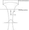
The Gaussian shear wave in a dispersive medium
- PMID: 24412170
- PMCID: PMC3943673
- DOI: 10.1016/j.ultrasmedbio.2013.10.023
The Gaussian shear wave in a dispersive medium
Abstract
In "imaging the biomechanical properties of tissues," a number of approaches analyze shear wave propagation initiated by a short radiation force push. Unfortunately, it has been experimentally observed that the displacement-versus-time curves for lossy tissues are rapidly damped and distorted in ways that can confound simple tracking approaches. This article addresses the propagation, decay and distortion of pulses in lossy and dispersive media, to derive closed-form analytic expressions for the propagating pulses. The theory identifies key terms that drive the distortion and broadening of the pulse. Furthermore, the approach taken is not dependent on any particular viscoelastic model of tissue, but instead takes a general first-order approach to dispersion. Examples with a Gaussian beam pattern and realistic dispersion parameters are given along with general guidelines for identifying the features of the distorting wave that are the most compact.
Keywords: Attenuation; Dispersion; Distortion; Propagation; Radiation force; Shear wave.
Copyright © 2014 World Federation for Ultrasound in Medicine & Biology. Published by Elsevier Inc. All rights reserved.
Figures


Similar articles
-
Comparison of fractional wave equations for power law attenuation in ultrasound and elastography.Ultrasound Med Biol. 2014 Apr;40(4):695-703. doi: 10.1016/j.ultrasmedbio.2013.09.033. Epub 2014 Jan 13. Ultrasound Med Biol. 2014. PMID: 24433745
-
A new method for shear wave speed estimation in shear wave elastography.IEEE Trans Ultrason Ferroelectr Freq Control. 2015 Dec;62(12):2106-14. doi: 10.1109/TUFFC.2015.007282. IEEE Trans Ultrason Ferroelectr Freq Control. 2015. PMID: 26670851
-
Physical models of tissue in shear fields.Ultrasound Med Biol. 2014 Apr;40(4):655-74. doi: 10.1016/j.ultrasmedbio.2013.11.001. Ultrasound Med Biol. 2014. PMID: 24582297 Review.
-
Error in estimates of tissue material properties from shear wave dispersion ultrasound vibrometry.IEEE Trans Ultrason Ferroelectr Freq Control. 2009 Apr;56(4):748-58. doi: 10.1109/TUFFC.2009.1097. IEEE Trans Ultrason Ferroelectr Freq Control. 2009. PMID: 19406703 Free PMC article.
-
Shear wave elastography of the liver - review on normal values.Z Gastroenterol. 2017 Feb;55(2):153-166. doi: 10.1055/s-0042-117226. Epub 2017 Feb 13. Z Gastroenterol. 2017. PMID: 28192849 Review. English.
Cited by
-
Shear wave propagation in viscoelastic media: validation of an approximate forward model.Phys Med Biol. 2019 Jan 8;64(2):025008. doi: 10.1088/1361-6560/aaf59a. Phys Med Biol. 2019. PMID: 30524099 Free PMC article.
-
Spatial localization of mechanical excitation affects spatial resolution, contrast, and contrast-to-noise ratio in acoustic radiation force optical coherence elastography.Biomed Opt Express. 2019 Oct 24;10(11):5877-5904. doi: 10.1364/BOE.10.005877. eCollection 2019 Nov 1. Biomed Opt Express. 2019. PMID: 31799053 Free PMC article.
-
An analytic, Fourier domain description of shear wave propagation in a viscoelastic medium using asymmetric Gaussian sources.J Acoust Soc Am. 2015 Aug;138(2):1012-22. doi: 10.1121/1.4927492. J Acoust Soc Am. 2015. PMID: 26328717 Free PMC article.
-
Plane-Wave Imaging Improves Single-Track Location Shear Wave Elasticity Imaging.IEEE Trans Ultrason Ferroelectr Freq Control. 2018 Aug;65(8):1402-1414. doi: 10.1109/TUFFC.2018.2842468. Epub 2018 Jun 1. IEEE Trans Ultrason Ferroelectr Freq Control. 2018. PMID: 29993543 Free PMC article.
-
Characterization of Viscoelastic Materials Using Group Shear Wave Speeds.IEEE Trans Ultrason Ferroelectr Freq Control. 2018 May;65(5):780-794. doi: 10.1109/TUFFC.2018.2815505. IEEE Trans Ultrason Ferroelectr Freq Control. 2018. PMID: 29733281 Free PMC article.
References
-
- Abramowitz M, Stegun IA. Handbook of mathematical functions with formulas, graphs, and mathematical tables. Vol. 364. Washington,:U.S.: Govt. Print. Off.; p. 1964.
-
- Baddour N. Multidimensional wave field signal theory: mathematical foundations. AIP Advances. 2011;1:0221201–02212024.
-
- Bercoff J, Tanter M, Fink M. Supersonic shear imaging: a new technique for soft tissue elasticity mapping. IEEE Trans Ultrason Ferroelectr Freq Control. 2004a;51:396–409. - PubMed
-
- Bercoff J, Tanter M, Muller M, Fink M. The role of viscosity in the impulse diffraction field of elastic waves induced by the acoustic radiation force. Ieee T Ultrason Ferr. 2004b;51:1523–36. - PubMed
Publication types
MeSH terms
Grants and funding
LinkOut - more resources
Full Text Sources
Other Literature Sources

