. 2014 Jan 20;24(2):156-164.
doi: 10.1016/j.cub.2013.12.018. Epub 2014 Jan 9.

INF2-mediated severing through actin filament encirclement and disruption

Affiliations

INF2-mediated severing through actin filament encirclement and disruption

Pinar S Gurel et al. Curr Biol. .

Abstract

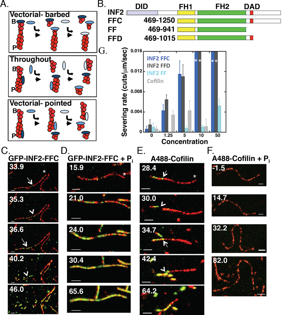
Background: INF2 is a formin protein with the unique ability to accelerate both actin polymerization and depolymerization, the latter requiring filament severing. Mutations in INF2 lead to the kidney disease focal segmental glomerulosclerosis (FSGS) and the neurological disorder Charcot-Marie Tooth disease (CMTD).

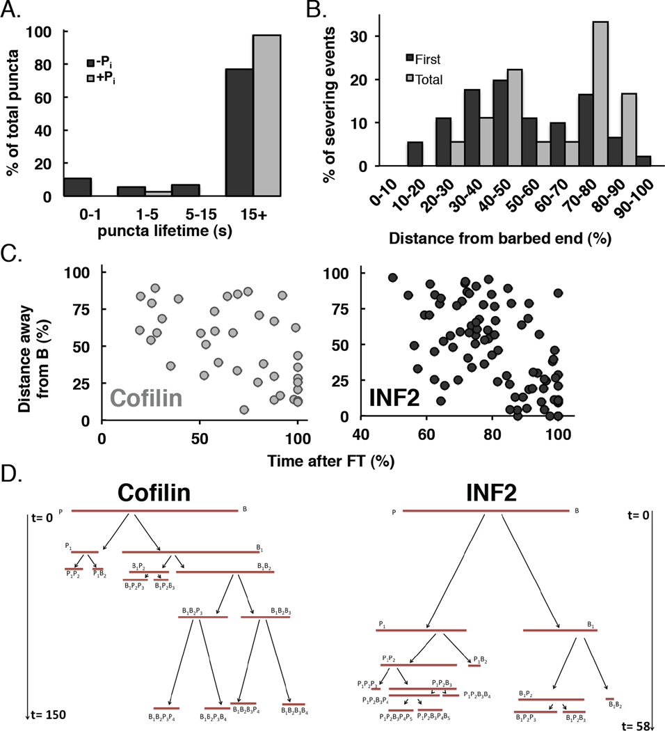
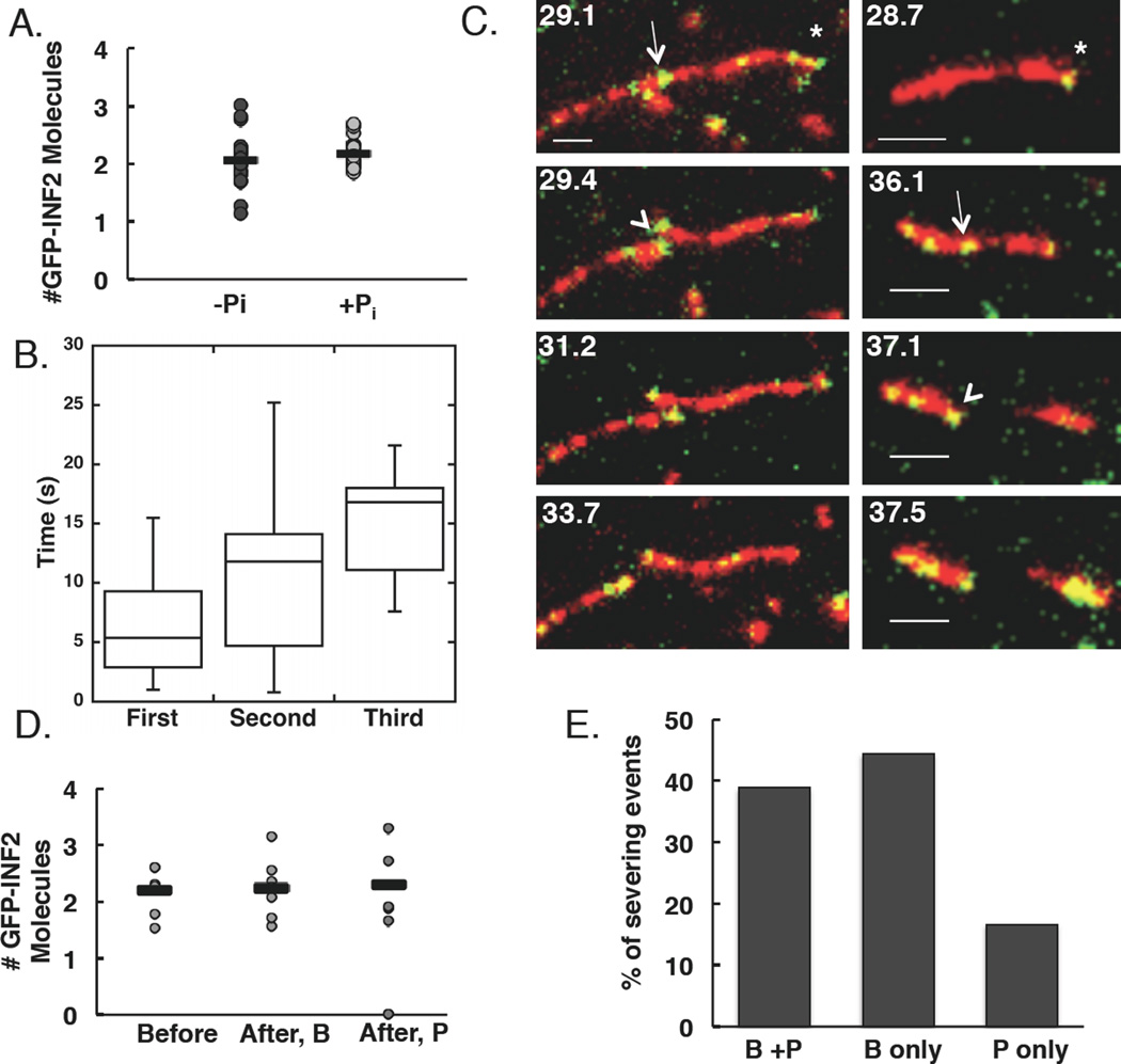
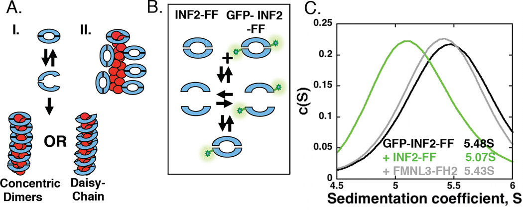
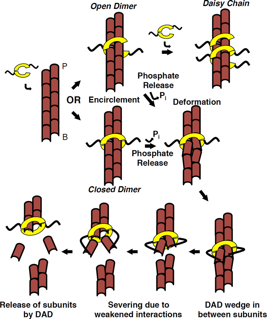
Results: Here, we compare the severing mechanism of INF2 with that of the well-studied severing protein cofilin. INF2, like cofilin, binds stoichiometrically to filament sides and severs in a manner that requires phosphate release from the filament. In contrast to cofilin, however, INF2 binds ADP and ADP-Pi filaments equally well. Furthermore, two-color total internal reflection fluorescence (TIRF) microscopy reveals that a low number of INF2 molecules, as few as a single INF2 dimer, are capable of severing, while measurable cofilin-mediated severing requires more extensive binding. Hence, INF2 is a more potent severing protein than cofilin. While a construct containing the FH1 and FH2 domains alone has some severing activity, addition of the C-terminal region increases severing potency by 40-fold, and we show that the WH2-resembling DAD motif is responsible for this increase. Helical 3D reconstruction from electron micrographs at 20 Å resolution provides a structure of filament-bound INF2, showing that the FH2 domain encircles the filament.

Conclusions: We propose a severing model in which FH2 binding and phosphate release causes local filament deformation, allowing the DAD to bind adjacent actin protomers, further disrupting filament structure.


Conflict of interest statement

The authors do not have any financial conflict of interest associated with this work.

Figures

Figure 1
Figure 1. INF2 severs filaments throughout their length
(A)Three models for INF2 severing. I) Vectorial-barbed: INF2 binds barbed end and removes a short filament fragment from that end. II) Throughout: INF2 binds and severs at random positions along filament side, creating two longer fragments. INF2 subsequently binds the newly created barbed end. III) Vectorial-pointed: INF2 binds and severs at ADP-actin protomers near pointed end. B, barbed. P, pointed. (B) Bar diagram showing human INF2 constructs (lengths in amino acids). DID-Diaphanous Inhibitory Domain, FH1- Formin Homology 1, FH2- Formin Homology 2, DAD- Diaphanous Autoregulatory Domain. (C) Two-color simultaneous TIRF microscopy montage of GFP-INF2-FFC (10nM) binding actin filament sides (1µM, 20% TAMRA initially) and severing on DDS/F127 treated slides. Time indicates seconds after INF2 addition (and wash-out of actin monomers). Asterisk indicates barbed end, arrow indicates GFP punctum correlated with severing, arrowhead indicates severing event. Scale bar 2 µm. Corresponds to Movie 1. (D)As in (C) but with GFP-INF2-FFC (100nM) + 10mM phosphate. Corresponds to Movie 2. (E) As in (C) but with A488-Cofilin (5nM). Corresponds to Movie 3. (F) As in (C) but with A488-cofilin (100nM) + 10mM phosphate. Corresponds to Movie 4. (G)Concentration dependence of severing rate for INF2-FFC, INF2-FFD, INF2-FF, and cofilin on DDS/F127 treated slides. N values: 18, 50, 48 severing events (7, 6, 6 filaments) for 0, 1.25, and 5nM INF2-FFC (dark blue); 15, 29, 40 events (6, 5, 7 filaments) for 0, 1.25, and 5nM INF2-FFD (dark grey); 9, 14, 15, 23 events (5, 6, 5, 5 filaments) for 0, 5, 10, and 50nM INF2-FF (light blue); 18, 42, 21, 4 events (7, 7, 5, 7 filaments) for 0, 1.25, 5, and 10nM cofilin (light grey). For 10 and 50 nM INF2-FFC and INF2-FFD, 10 and 6 filaments were examined respectively, but severing events were too rapid to quantify (depicted by dark blue or dark grey bar to the maximum rate). The highest concentrations tested (1 µM INF2-FFC and 500nM INF2-FFD) produce similar results.
Figure 2
Figure 2. INF2 binds throughout the filament but severs towards pointed end
(A)Lifetimes of filament-bound puncta in (A). >15s represents puncta that did not dissociate during the observation period (180 sec). n=74 and 39 puncta (13 and 6 filaments) in the absence and presence of phosphate respectively. (B)Histogram of severing positions for GFP-INF2-FFC (5–20nM) on DDS/F127 treated slides. 18 filaments (91 severing events) quantified (average length 12.3µm, range 5.83–24.22µm at time = 0). Barbed end fragments were measured as a percentage of total filament length. (C) Correlation of time of severing with respect to distance from original barbed end (as % of original length). n=80 and 38 severing events (11 and 7 filaments) for GFP-INF2-FFC and A488-cofilin, respectively. Filaments were12.5 and 7.86µm mean length (range 6.49–21.59 and 4.55–11.05µm) prior to INF2-FFC or cofilin addition respectively. Severing events were observed for 57.5–112.3 and 68.6–170.8 s after addition of INF2 or cofilin respectively. Note x-axis scale is different between INF2 and cofilin. (D)Tree map diagraming severing events on individual filaments with time (sec) on the y-axis. t=0 denotes time of cofilin or INF2-FFC addition. B and P denote barbed and pointed ends. Severed filament fragments are named based on severing history with subscripts denoting lineage (B1 being the first barbed end fragment, B1B2 being the second barbed end fragment originating from the first barbed end fragment, and so forth). Original filament lengths: 11.1 (cofilin) and 13.6 µm (INF2). Time and sizes drawn to scale. Additional examples in Figure S2.
Figure 3
Figure 3. INF2 binds filament sides as single dimers and severs at binding sites
(A)Number of GFP-INF2-FFC molecules localized at diffraction-limited puncta (n=28 and 25 puncta (8 and 6 filaments) in the absence and presence of phosphate respectively). Averages (bars) are 2.06 and 2.17 molecules in the absence and presence of phosphate respectively. (B) Box and whisker quantification of severing time after punctum binding for first, second, and third severing events. Line indicates mean, box represents values 25–75% of totals, whiskers indicate full range of values. n= 14, 9, and 5 events. (C) Two examples of severing at sites where an apparent single GFP-INF2-FFC dimer (green) is bound to TAMRA-actin (red). Asterisk indicates barbed end, arrow indicates punctum correlated with severing, arrowhead indicates severing event. Left panels: GFP-INF2 puncta present at both new barbed and pointed ends after severing. In right panels: GFP-INF2 punctum only at new barbed end after severing. Scale bar 2 µm. Corresponds to Movies 5 and 6. (D)Distribution of GFP-INF2-FFC localization after severing for events occurring at sites where two GFP-INF2-FFC were located prior to severing. After, B = new barbed end. After, P = new pointed end. Bars represent averages from 7 events. Note: two events result in punctum localization at only new barbed end. (E) Localization of puncta after a severing event with respect to newly created barbed or pointed ends. All severing events included, regardless of the number of GFP-INF2-FFC molecules present before the event. n=18 filaments.
Figure 4
Figure 4. INF2 is a dissociating dimer that binds stoichiometrically to filament sides
(A)Two models for INF2 binding filament sides. I. FH2 dimer partially dissociates in order to encircle the filament. Encirclement could occur either as concentric dimers or in a daisy-chain, where the lasso-region of one FH2 domain interacts with the post region of an adjacent FH2. II. FH2 dimer binds using interactions between the exterior of the dimer and the filament side. Two possible side-binding orientations shown, as well as a barbed end-bound dimer. (B) Schematic representation of heterogenous dimer formation in velocity analytical ultracentrifucation experiments. If dimer dissociation is appreciable, a heterogeneous dimer should form upon mixing GFP-labeled and unlabeled FH2 domains, resulting in a shift in S value for the GFP signal. (C)Velocity analytical ultracentrifugation profiles of GFP-INF2-FF (2µM) alone or mixed with unlabeled INF2-FF (20µM) or FMNL3-FH2 (20µM).
Figure 5
Figure 5. Examination of INF2-FFC binding actin filaments by electron microscopy
A) 3D reconstructions from negative staining EM images of bare (left) and INF2-FFC bound (right) actin filaments. Barbed (B) and pointed (P) ends indicated. B) Zoomed images of full actin/INF2-FFC reconstruction (inner and outer densities) with actin monomer (PDB: 3MFP, pink ribbon) on inner density alone (left panel) or on INF2-bound filaments with FH2 crystal backbone ribbon (PDB 4EAH, green ribbon). Note density unaccounted for by either actin or FH2 (arrow). Three angles shown. C) Schematic model of FH2 (green, blue) binding to filament (red, yellow), based on structural data in agreement with color scheme of [34].
Figure 6
Figure 6. Model of INF2 severing mechanism
INF2 dimer partially dissociates, then encircles the filament, either as a closed ring or an open dimer that can form a daisy-chain. Subsequent steps shown in the closed ring conformation, but could also occur as the open dimer. INF2 binding causes a change in helical rise. Upon phosphate release, INF2 causes further local filament deformation, partially exposing the DAD binding site between subdomains 1 and 3 of adjacent actin protomers. DAD binding destabilizes the filament by wedging between protomers. These destabilizations lead to severing at this weak point. Upon severing, INF2 remains bound to the new barbed end, and DAD releases bound actin monomers removed from filament. Our data suggest that DAD binding to the barbed end side of the FH2 is favored.

Similar articles

Cited by

References

    1. Chhabra ES, Higgs HN. The many faces of actin: matching assembly factors with cellular structures. Nat Cell Biol. 2007;9:1110–1121. - PubMed
    1. Moseley JB, Sagot I, Manning AL, Xu Y, Eck MJ, Pellman D, Goode BL. A conserved mechanism for Bni1- and mDia1-induced actin assembly and dual regulation of Bni1 by Bud6 and profilin. Mol Biol Cell. 2004;15:896–907. - PMC - PubMed
    1. Chesarone MA, Goode BL. Actin nucleation and elongation factors: mechanisms and interplay. Curr Opin Cell Biol. 2009;21:28–37. - PMC - PubMed
    1. Paul AS, Pollard TD. Review of the mechanism of processive actin filament elongation by formins. Cell Motil Cytoskeleton. 2009;66:606–617. - PMC - PubMed
    1. Higgs HN, Peterson KJ. Phylogenetic analysis of the formin homology 2 domain. Mol Biol Cell. 2005;16:1–13. - PMC - PubMed

Publication types

Substances