. 2014 Jan 16;40(1):25-39.
doi: 10.1016/j.immuni.2013.10.021. Epub 2014 Jan 9.

Induction of innate lymphoid cell-derived interleukin-22 by the transcription factor STAT3 mediates protection against intestinal infection

Affiliations

Induction of innate lymphoid cell-derived interleukin-22 by the transcription factor STAT3 mediates protection against intestinal infection

Xiaohuan Guo et al. Immunity. .

Abstract

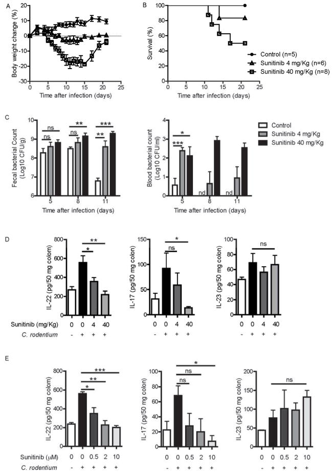
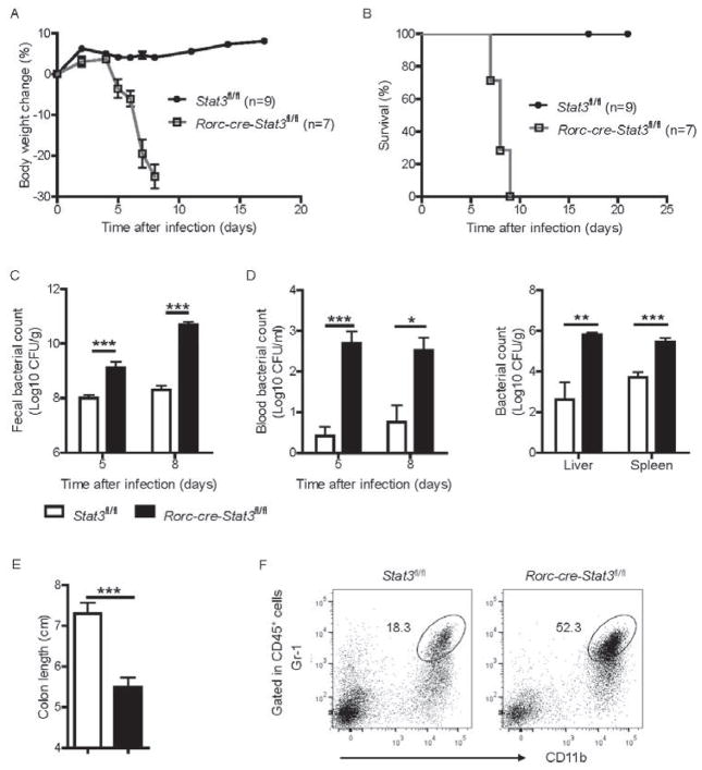
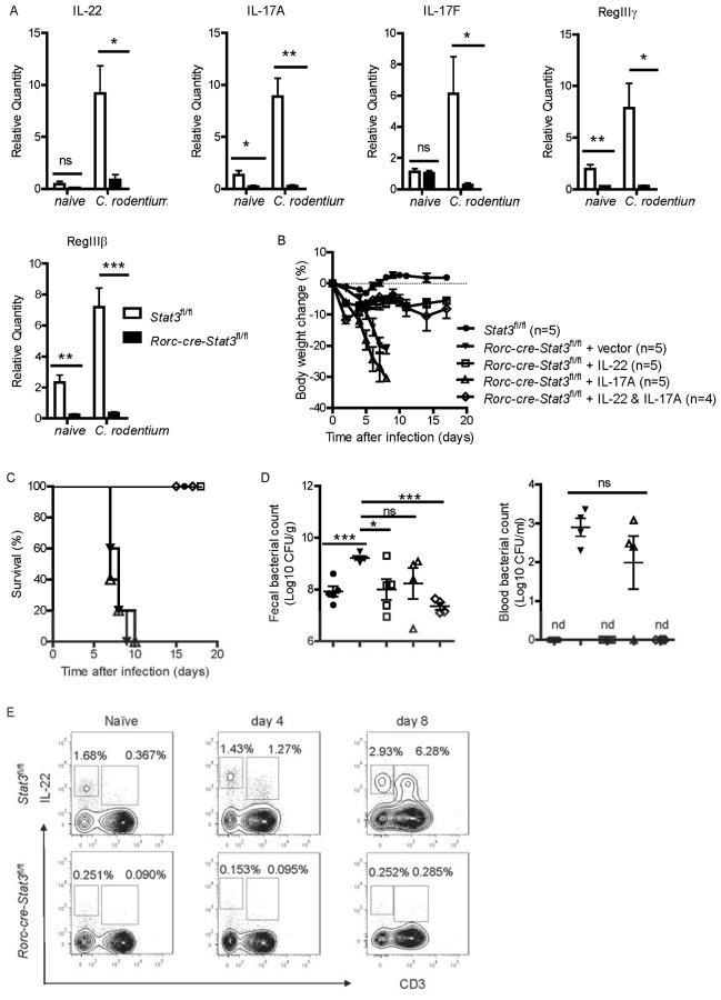
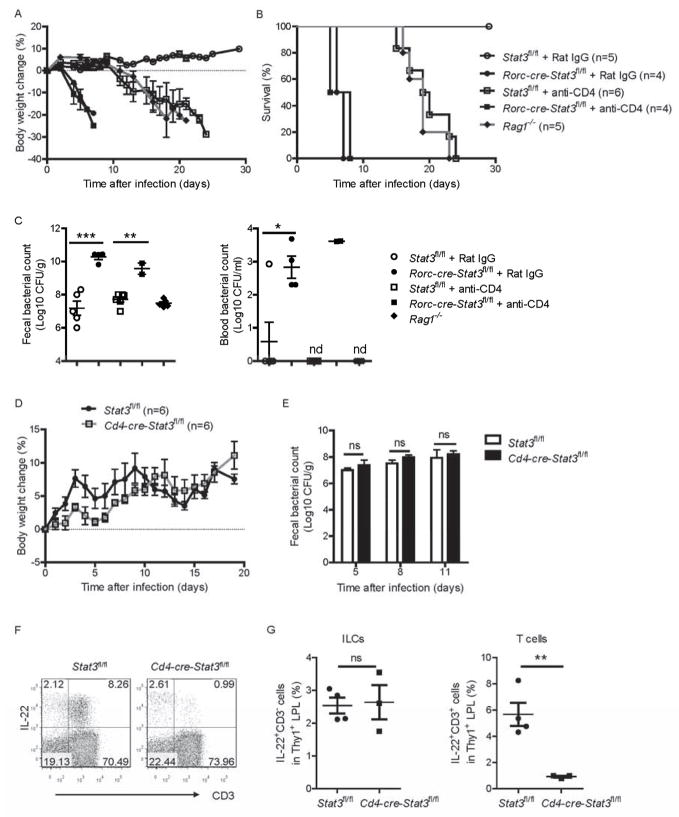
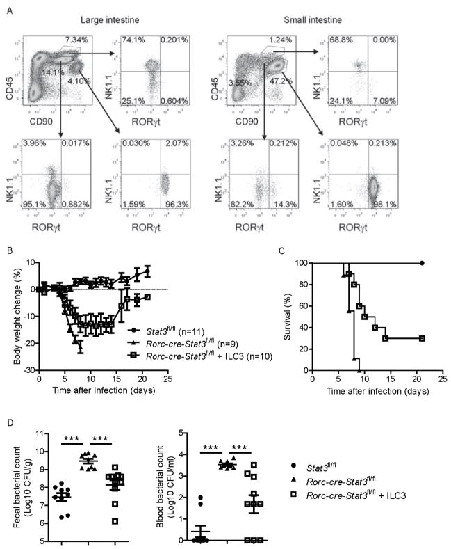
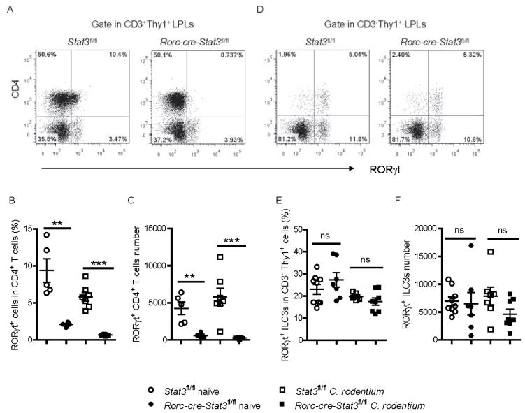
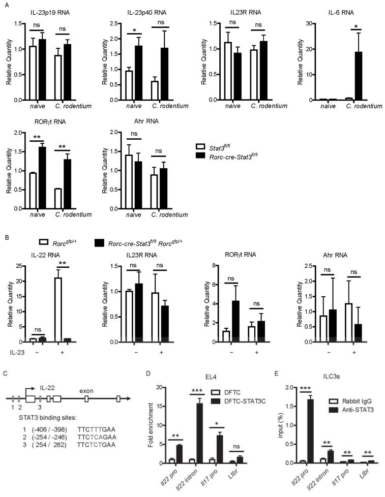
Inhibitors of the transcription factor STAT3 target STAT3-dependent tumorigenesis but patients often develop diarrhea from unknown mechanisms. Here we showed that STAT3 deficiency increased morbidity and mortality after Citrobacter rodentium infection with decreased secretion of cytokines including IL-17 and IL-22 associated with the transcription factor RORγt. Administration of the cytokine IL-22 was sufficient to rescue STAT3-deficient mice from lethal infection. Although STAT3 was required for IL-22 production in both innate and adaptive arms, by using conditional gene-deficient mice, we observed that STAT3 expression in RORγt(+) innate lymphoid cells (ILC3s), but not T cells, was essential for the protection. However, STAT3 was required for RORγt expression in T helper cells, but not in ILC3s. Activated STAT3 could directly bind to the Il22 locus. Thus, cancer therapies that utilize STAT3 inhibitors increase the risk for pathogen-mediated diarrhea through direct suppression of IL-22 from gut ILCs.


Figures

Figure 1
Figure 1. Sunitinib impairs the host defense against intestinal C. rodentium infection
(A–C) 8 weeks old wild type mice were orally inoculated with 2 × 10⁹ CFU of C. rodentium. Sunitinib (4 mg/Kg, n=6 or 40 mg/Kg, n=8) or control (n=5) was administered orally from day 0 to day 7, once daily. Body weight was measured at the indicated time points (A) and survival rates (B) are shown. (C) Bacterial titers from blood and fecal homogenate cultures at indicated day post infection in the mice in A, B. (D) IL-22, IL-17 and IL23 levels from the colon culture supernatants of Sunitinib or control treated mice at day 5 after infection (n=5 each group). (E) The colons from untreated mice (n=3) were removed at day 5 after infection, and cultured with Sunitinib at indicated concentrations for 24 hours in vitro. IL-22, IL-17 and IL23 levels were examined in the colon culture supernatants. *P<0.05, **P<0.01, ***P<0.001; ns, no significant difference (Student’s t-test); nd, nondetectable. Data are representative of two independent experiments (mean ± s.e.m.). See also Figure S1.
Figure 2
Figure 2. STAT3 signaling is essential for control of intestinal C. rodentium infection
(A–F) 7 weeks old Rorc-cre-Stat3fl/fl (n = 7) and their littermate wild type Stat3fl/fl (n = 9) mice were orally inoculated with C. rodentium (2 × 10⁹ CFU) and body weight was measured at the indicated time points. Body weight change (A) and survival rates (B) are shown. (C) Bacterial titers from fecal homogenate cultures at indicated day post infection in the mice in A, B. (D) Bacterial titers from blood and spleen, liver homogenate cultures at indicated day post infection are shown. (E) Rorc-cre-Stat3fl/fl mice show colon shortening at day 8 post infection. (F) Rorc-cre-Stat3fl/fl mice show more inflammatory monocytes or neutrophils infiltration in the colon. LPLs were isolated from colon and gated in CD45+ cells. *P<0.05, **P<0.01, ***P<0.001 (Student’s t-test). Data are representative of three independent experiments (A–C; mean ± s.e.m.) or two independent experiments (D–E; mean ± s.e.m.). See also Figure S2.
Figure 3
Figure 3. IL-22, but not IL-17, is an essential and sufficient pathway downstream of STAT3 signaling to protect mice against C. rodentium infection
(A) Rorc-cre-Stat3fl/fl and wild type Stat3fl/fl littermate mice (n = 5 per group) were infected with 2 × 10⁹ CFU of C. rodentium. The mRNA expression of IL-22, IL-17A, IL-17F and IL-22- dependent RegIIIγ and RegIIIβ antimicrobial proteins in the colon of naïve or 5 days infected mice were measured by real-time. *P<0.05, **P<0.01, ***P<0.001 (Student’s t-test). Data are representative of two independent experiments (A; mean ± s.e.m.). (B–D) Stat3fl/fl (n = 5) and Rorc-cre-Stat3fl/fl mice were orally inoculated with 2 × 10⁹ CFU of C. rodentium. 10 μg control vector (n = 5), IL-17 (n = 5), IL-22 (n = 5), or both IL-17 and IL-22 (n = 4) expressing plasmids were hydrodynamic injected into Rorc-cre-Stat3fl/fl mice through the tail vein 6 hr after C. rodentium infection. Body weight changes (B) and survival rates (C) were monitored at indicated time points. (D) CFU counts of C. rodentium in the fecal pellets and blood on day 5 after infection are shown. Data are representative of two independent experiments (mean ± s.e.m.). *P<0.05, ***P<0.001; ns, no significant difference (Student’s t-test). nd, nondetectable. (E) STAT3 signaling is required for both innate and adaptive IL-22 production. IL-22 expression in CD3 and CD3+ cells were analyzed by intracellular cytokine staining followed by flow cytometry. Rorc-cre-Stat3fl/fl and wild type Stat3fl/fl littermate mice were infected with 2 × 10⁹ CFU of C. rodentium. Intestinal LPLs were isolated from the colons of Stat3fl/fl or Rorc-cre-Stat3fl/fl mice at day 4 and day 8 post-infection. Cells were stimulated with IL-23 (25 ng/ml), PMA (50 ng/ml) and ionomycin (750 ng/ml) for 4 hours and gated in Thy1+ lymphocytes. Naïve mice were used as control. Data are representative of three independent experiments. See also Figure S3.
Figure 4
Figure 4. STAT3 signaling and IL-22 from RORγt+ ILCs, but not T cells, are essential for the protection from C. rodentium infection
(A–C) Rorc-cre-Stat3fl/fl and wild type Stat3fl/fl mice were infected with 5 × 10⁶ CFU of C. rodentium and injected intraperitoneally with mAb GK1.5 or Rat IgG (50 μg per mouse each time) at day −5, 0, 5, 10 and 15 post infection for depletion of CD4+ T cells (4 to 6 mice per group). Average body weight change (A) and survival rates (B) at the indicated time points are shown. (C) Bacterial titers from blood and fecal homogenate cultures at day 5 post infection. Each dot represents one individual mouse. (D, E) Cd4-cre-Stat3fl/fl (n=6) and wild type Stat3fl/fl mice (n=6) were orally infected with 2 × 10⁹ CFU of C. rodentium. (D) Body weight changes are shown at the indicated time points. (E) Bacterial titers from fecal homogenate cultures at indicated day post infection in the mice in D. (F, G) IL-22 expression in CD3 and CD3+ cells were analyzed by intracellular cytokine staining followed by flow cytometry. Cd4-cre-Stat3fl/fl and wild type Stat3fl/fl littermate mice were infected with 2 × 10⁹ CFU of C. rodentium. LPLs were isolated from the colons at day 8 post-infection. Cells were stimulated with IL-23 (25 ng/ml), PMA (50 ng/ml) and ionomycin (750 ng/ml) for 4 hours and gated in Thy1+ lymphocytes. Each dot represents one individual mouse. *P<0.05, **P<0.01, ***P<0.001; ns, no significant difference (Student’s t-test). nd, nondetectable. Data are representative of two independent experiments (A–G; mean ± s.e.m.). See also Figure S4.
Figure 5
Figure 5. CD90hiCD45lo ILC3s protect Rorc-cre-Stat3fl/fl mice from C. rodentium infection
(A) Intestinal CD90hiCD45lo LPLs are RORγt+ ILCs. LPLs were isolated from both the large intestine and small intestine of Rag1−/− mice and gated by CD45 and CD90 expression. Three separate populations, CD90hiCD45lo, CD90loCD45int and CD90loCD45hi were further analyzed with the expression of NK1.1 and RORγt. Data are representative of two independent experiments. (B–D) Rag1−/− mice were infected with 2 × 10⁹ CFU of C. rodentium and CD90hiCD45lo ILC3s were sorted from both the colon and small intestine LPLs at day 1 post infection. Sorted ILC3s were stimulated with IL-23 (25 ng/ml) for 1 hour, then were transferred by i.v. injection (2 × 10⁵ cells per mouse) into Rorc-cre-Stat3fl/fl recipients at day 0 and day 3 post inoculated with 5 × 10⁶ CFU of C. rodentium. Untreated Rorc-cre-Stat3fl/fl and wild type Stat3fl/fl littermate mice were as control. Body weight changes (B) and survival rates (C) were monitored at indicated time points. (D) CFU counts of C. rodentium in the fecal pellets and blood on day 5 after infection are shown. Data are pooled from two independent experiments with nine to eleven mice per group (mean ± s.e.m.). ***P<0.001 (Student’s t-test). See also Figure S5.
Figure 6
Figure 6. The development of RORγt+ T helper cells, but not RORγt+ ILCs is dependent on STAT3 signaling
(A–F) Colonic LPLs were isolated from Stat3fl/fl and Rorc-cre-Stat3fl/fl littermate mice before or 5 days after C. rodentium infection. (A, D) RORγt and CD4 expression were analyzed in colonic LPLs from naïve mice by flow cytometry after gating on Thy1+ CD3+ adaptive lymphocytes and Thy1+ CD3 innate lymphoid cells. (B, C) Percentage of RORγt+ cells in the CD4+ CD3+ T cell population, as well as the absolute numbers of RORγt+ CD4+ T cells in the colons of naïve and C. rodentium infected Stat3fl/fl and Rorc-cre-Stat3fl/fl mice are shown. (E, F) Percentage of RORγt+ cells in the CD3 Thy1+ ILCs population, as well as the absolute numbers of RORγt+ ILCs in the colons of naïve and C. rodentium infected Stat3fl/fl and Rorc-cre-Stat3fl/fl mice are shown. **P<0.01, ***P<0.001; ns, no significant difference (Student’s t-test). Data are representative of three independent experiments (mean ± s.e.m.). Each dot represents one individual mouse (B, C, E, F). See also Figure S6.
Figure 7
Figure 7. STAT3 directly binds to the Il22 locus and regulates IL-22 production
(A) Rorc-cre-Stat3fl/fl and Stat3fl/fl littermate mice (n = 5 per group) were infected with 2 × 10⁹ CFU of C. rodentium. The mRNA expression of IL-23 p19, IL-23 p40, IL23R, IL-6, RORγt and Ahr in the colon were measured by real-time PCR before and 5 days after infection. Data are representative of two independent experiments (mean ± s.e.m.). (B) RORγt+ ILCs were purified by flow cytometric sorting from colonic LPLs of Rorc-cre-Stat3fl/fl Rorcgfp/+ and Rorcgfp/+ mice and stimulated with or without IL-23 (25 ng/ml) for 1 hour. The mRNA expression of IL-22, IL-23R, RORγt and Ahr were measured by real-time PCR. Data are representative of two independent experiments (mean ± s.e.m.). (C) STAT3 binding sites at the Il22 locus are shown. (D) STAT3 binding at the Il22 locus in EL4 stable cell lines stably expressing either flag peptide (DFTC) or flag-tagged STAT3C (DFTC-STAT3C) was monitored using a ChIP assay. The anti-flag antibody immunoprecipitates were analyzed by real-time PCR. The fold enrichment of STAT3C binding at each locus was normalized to DFTC-empty EL4 cells. Ltbr and Il17 loci were used as negative and positive controls in the ChIP assays, respectively. Data are representative of three independent experiments (mean ± s.e.m. of triplicate samples of real-time PCR). (E) CD90hiCD45lo ILC3s were sorted from Rag1−/− mice and stimulated with IL-23 (25 ng/ml) for 30 minutes. ChIP assays with anti-STAT3 mAb showed enhanced enrichment of STAT3 at the Il22 locus in primary ILC3s. Ltbr loci lacking STAT3 binging sites was used as negative controls. Data are representative of two independent experiments (mean ± s.e.m. of quadruplicate samples of real-time PCR). *P<0.05, **P<0.01, ***P<0.001; ns, no significant difference (Student’s t-test).

Similar articles

Cited by

References

    1. Basu R, O’Quinn DB, Silberger DJ, Schoeb TR, Fouser L, Ouyang W, Hatton RD, Weaver CT. Th22 cells are an important source of IL-22 for host protection against enteropathogenic bacteria. Immunity. 2012;37:1061–1075. - PMC - PubMed
    1. Bry L, Brenner MB. Critical role of T cell-dependent serum antibody, but not the gut-associated lymphoid tissue, for surviving acute mucosal infection with Citrobacter rodentium, an attaching and effacing pathogen. J Immunol. 2004;172:433–441. - PubMed
    1. Bry L, Brigl M, Brenner MB. CD4+-T-cell effector functions and costimulatory requirements essential for surviving mucosal infection with Citrobacter rodentium. Infect Immun. 2006;74:673–681. - PMC - PubMed
    1. Eberl G. Development and evolution of RORgammat+ cells in a microbe’s world. Immunol Rev. 2012;245:177–188. - PubMed
    1. Eberl G, Littman DR. Thymic origin of intestinal alphabeta T cells revealed by fate mapping of RORgammat+ cells. Science. 2004;305:248–251. - PubMed

Publication types

MeSH terms