. 2014 Jan 16;40(1):153-65.
doi: 10.1016/j.immuni.2013.11.021. Epub 2014 Jan 9.

Proatherogenic conditions promote autoimmune T helper 17 cell responses in vivo

Affiliations

Proatherogenic conditions promote autoimmune T helper 17 cell responses in vivo

Hoyong Lim et al. Immunity. .

Abstract

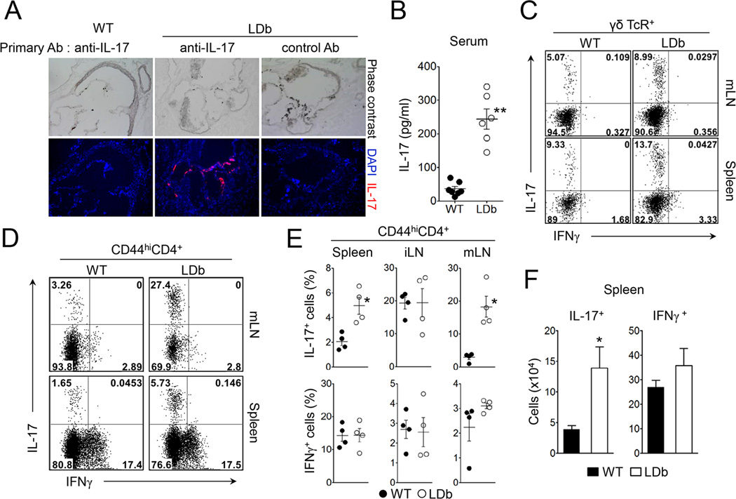
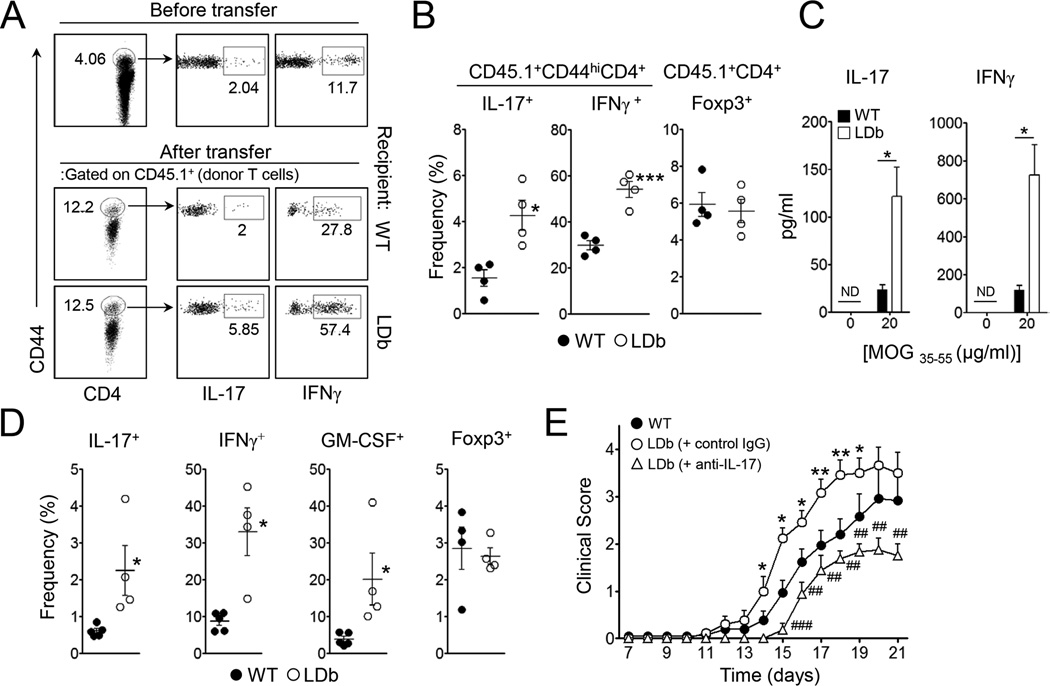
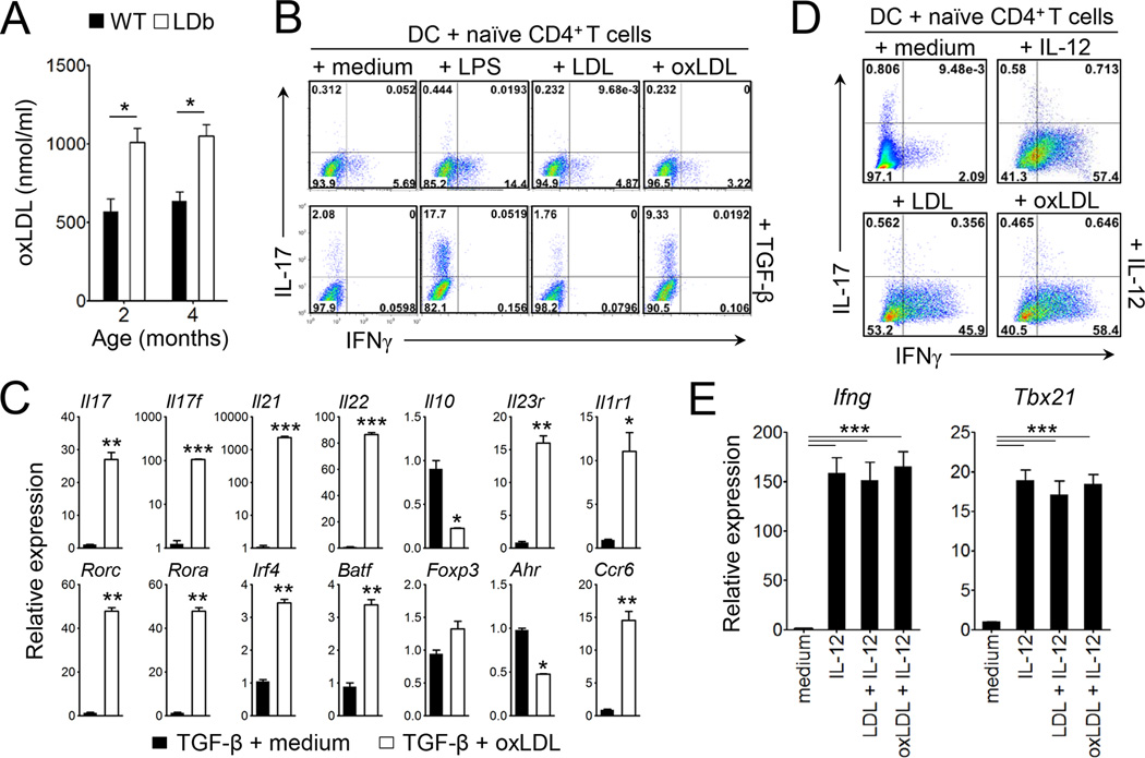
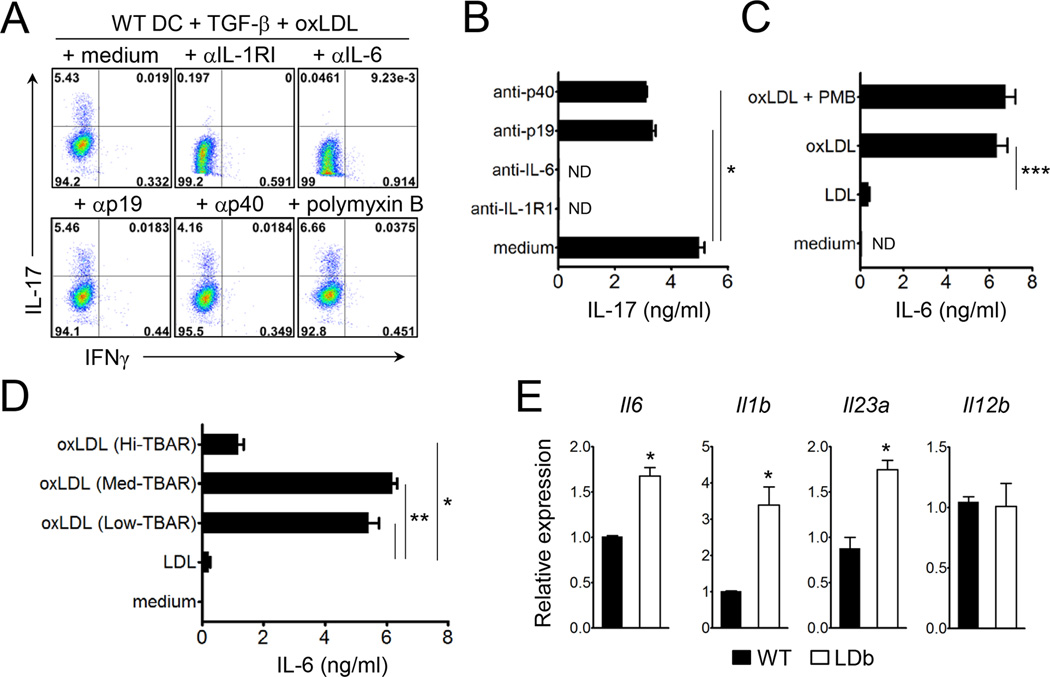
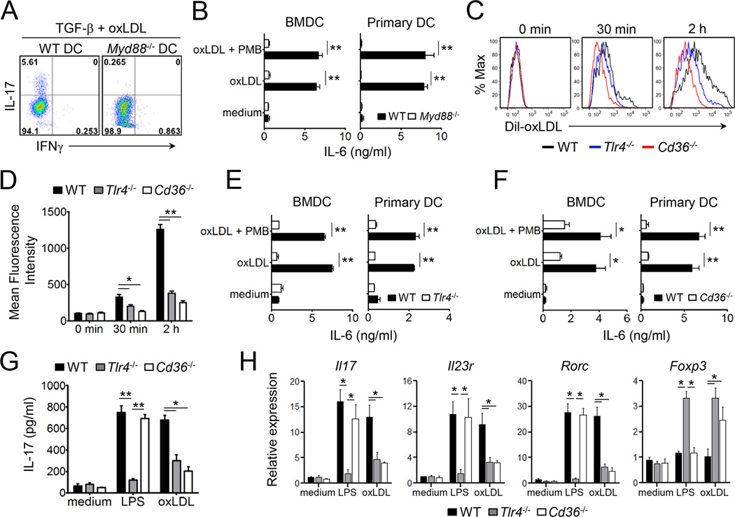
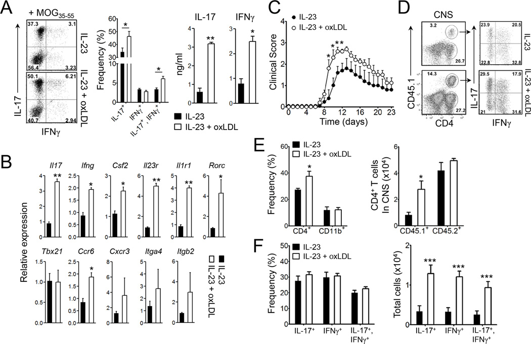
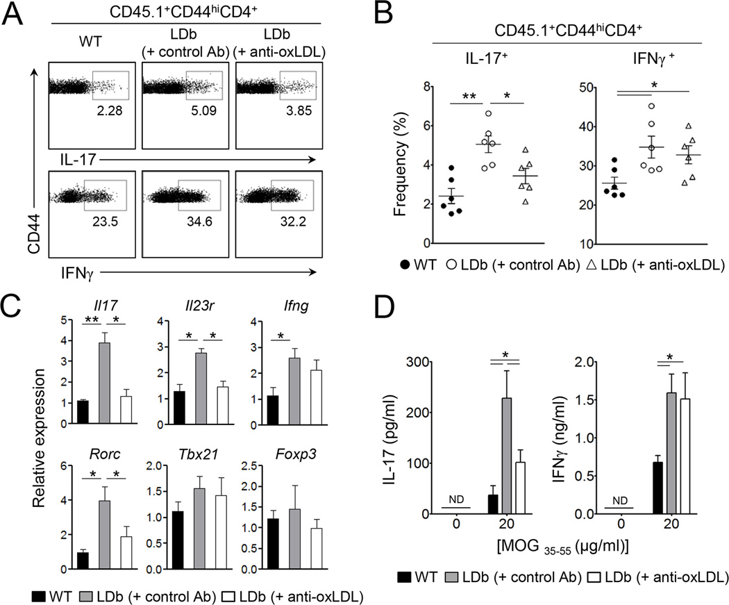
Patients with systemic autoimmune diseases show increased incidence of atherosclerosis. However, the contribution of proatherogenic factors to autoimmunity remains unclear. We found that atherogenic mice (herein referred to as LDb mice) exhibited increased serum interleukin-17, which was associated with increased numbers of T helper 17 (Th17) cells in secondary lymphoid organs. The environment within LDb mice was substantially favorable for Th17 cell polarization of autoreactive T cells during homeostatic proliferation, which was considerably inhibited by antibodies directed against oxidized low-density lipoprotein (oxLDL). Moreover, the uptake of oxLDL induced dendritic-cell-mediated Th17 cell polarization by triggering IL-6 production in a process dependent on TLR4, CD36, and MyD88. Furthermore, self-reactive CD4(+) T cells that expanded in the presence of oxLDL induced more profound experimental autoimmune encephalomyelitis. These findings demonstrate that proatherogenic factors promote the polarization and inflammatory function of autoimmune Th17 cells, which could be critical for the pathogenesis of atherosclerosis and other related autoimmune diseases.


Figures

Figure 1
Figure 1. LDb mice exhibit increased Th17 cell population
(A) The expression of IL-17 in aortic sinus in LDb or wild-type mice. Aortic sinus sections obtained from LDb or wild-type mice (4 month-old) were stained with isotype control or anti-IL- 17 polyclonal antibody (red) and DAPI (blue). (B) IL-17 levels in serum obtained from LDb or wild-type mice were measured (n=6). (C) IL-17 and IFNγ expression on γδTcR+ cells from the spleen or mesenteric lymph nodes of LDb and wild-type mice. (D and E) IL-17 and IFNγ expression on CD44hiCD4+ T cells from the spleen, inguinal lymph nodes (iLN) or mesenteric lymph nodes (mLN) of LDb and wild-type mice. (F) Absolute numbers of IL-17- and IFNγ-producing CD4+ T cells in the spleens (n=4). Data represent three independent experiments. Data shown are mean ± SEM. *, p<0.05; **, p<0.01 in comparison with wild-type mice. See also Figure S1 to S4.
Figure 2
Figure 2. LDb mice promote Th17 and Th1 cell responses of self-reactive CD4+ T cells in vivo
Total splenocytes from 2D2 mice (CD45.1+) were i.v. transferred into sublethally irradiated LDb or wild-type mice (CD45.2+, n=4). The recipients remained without further treatment (A–C), or were additionally immunized with MOG35–55 in CFA (D and E). (A and B) The frequencies of IL-17- or IFNγ-producers among CD44hi donor CD4+ T cells and Foxp3+ cells among donor CD4+ T cells in the spleens. (C) The amounts of IL-17 and IFNγ in the supernatant after 3 days culture of splenocytes with cognate peptide. (D) The frequencies of IL-17+, IFNγ+, GM-CSF+ or Foxp3+ cells among donor CD4+ T cells in the spleens 2 weeks after immunization. (E) Daily clinical scores of EAE (n=10–12). Data represent two independent experiments. Data shown are mean ± SEM. *, p<0.05; **, p<0.01; ***, p<0.001 in comparison with wild-type. ##, p<0.01; ###, p<0.001 in comparison with control IgG injected LDb mice.
Figure 3
Figure 3. Oxidized LDL promotes dendritic cell-mediated Th17 cell differentiation in vitro
(A) Concentration of oxLDL in serum at the age of 2 or 4 month of wild-type and LDb mice (n=4–5). (B–E) Purified naive CD4+ T cells were co-cultured with bone marrow-derived DC in the presence of soluble anti-CD3 under the indicated conditions for 4 days. (B and D) The frequencies of IL-17- or IFNγ-producers among CD4+ T cells. (C and E) The levels of mRNA transcripts of the indicated genes. Data are representative of at least three independent experiments. Data shown are mean ± SEM. *, p<0.05; **,p<0.01; ***,p<0.001. See also Figure S5 and S6.
Figure 4
Figure 4. Oxidized LDL induces the production of Th17 cell promoting cytokines
(A and B) Naive CD4+ T cells were co-cultured with bone marrow-derived DC under the indicated conditions in the presence or absence of indicated antibodies for 4 days. (A) The frequencies of IL-17- or IFNγ-producers among CD4+ T cells. (B) IL-17 production by CD4+ T cells after stimulation with plate-bound anti-CD3 (1 µg/ml). (C and D) IL-6 production by bone marrow-derived DCs after stimulation with 20 µg/ml LDL or oxLDL (Medium Tbar) (C), or with 20 µg/ml oxLDL with Hi-, Medium- or Low Tbars (D). (E) Relative mRNA expression of indicated genes in the freshly purified CD11c+ DCs from the spleens of LDb and wild-type mice. Data are representative of at least three independent experiments. Data shown are mean ± SEM. *, p<0.05; **, p<0.01; ***, p<0.001. ND, not detected.
Figure 5
Figure 5. TLR4, CD36, and MyD88 are essential for the oxLDL-mediated differentiation of Th17 cells
(A) The frequencies of IL-17- or IFNγ-producers among CD4+ T cells co-cultured with wild-type or Myd88−/− DC under the indicated conditions. (B) Production of IL-6 from freshly isolated DCs or bone marrow-derived DCs after stimulation with oxLDL (20 µg/ml). (C and D) The uptake of Dil-oxLDL by DC freshly isolated from the spleens of wild-type, Tlr4−/−, or Cd36−/− mice. (E and F) Production of IL-6 by freshly isolated or bone marrow-derived DCs from wild-type, Tlr4−/− (E) or Cd36−/− (F) mice (n=3–4) after stimulation with oxLDL. (G and H) Naïve CD4+ T cells were co-cultured with bone marrow-derived DCs from wild-type, Tlr4−/− or Cd36−/− mice plus soluble anti-CD3 and TGFβ in the presence or absence of LPS or oxLDL for 4 days. The production of IL-17 by T cells (G), and the levels of mRNA transcripts of the indicated genes (H). Data are representative of at least two independent experiments. Data shown are mean ± SEM. *, p<0.05; **, p<0.01.
Figure 6
Figure 6. MOG-reactive T cells expanded in the presence of oxLDL induces enhanced EAE
Lymphoid cells from the draining LNs of B6.SJL mice (CD45.1+) that were previously immunized with MOG35–55 in CFA were restimulated with the same peptide in the presence or absence of oxLDL and IL-23 for 5 days. (A) Production of IL-17 and IFNγ by CD4+ T cells and (B) mRNA levels of the indicated genes. (C–F) The ex vivo expanded MOG35–55-reactive CD4+ T cells (1×10⁶ cells/transfer) were i.v. transferred into congenic recipients (CD45.2+), which were then immunized with MOG35–55 in CFA (n=9–10). (C) The clinical severity of EAE. (D and F) the frequencies and absolute numbers of IL-17- or IFNγ-producing donor CD4+ T cells in the CNS tissues. (E) The proportion and absolute numbers of CD4+ and CD11b+ cells. Data are representative of three independent experiments. Data shown are mean ± SEM. *, p<0.05; **, p<0.01; ***, p<0.001. See also Figure S7.
Figure 7
Figure 7. Anti-oxLDL diminishes autoreactive Th17 cell responses in LDb mice
Total splenocytes from 2D2 mice (CD45.1+) were i.v. transferred into sublethally irradiated LDb or wild-type mice (CD45.2+, n=6) at day 0. LDb recipients were i.p. injected with anti-oxLDL or isotype control antibody at day -1, 1, and 3. (A and B) The frequencies of IL-17- or IFNγ-producers among CD44hi donor CD4+ T cells in the spleens. (C) Transcript levels of indicated genes. (D) The amounts of IL-17 and IFNγ in the supernatant after 3 days culture of splenocytes cells stimulated with MOG33–55 peptide. Data are representative of two independent experiments. Data shown are mean ± SEM. *, p<0.05; **, p<0.01.

Comment in

References

    1. Chawla A, Barak Y, Nagy L, Liao D, Tontonoz P, Evans RM. PPAR-gamma dependent and independent effects on macrophage-gene expression in lipid metabolism and inflammation. Nat Med. 2001;7:48–52. - PubMed
    1. Chung Y, Chang SH, Martinez GJ, Yang XO, Nurieva R, Kang HS, Ma L, Watowich SS, Jetten AM, Tian Q, Dong C. Critical regulation of early Th17 cell differentiation by interleukin-1 signaling. Immunity. 2009;30:576–587. - PMC - PubMed
    1. Cui G, Qin X, Wu L, Zhang Y, Sheng X, Yu Q, Sheng H, Xi B, Zhang JZ, Zang YQ. Liver X receptor (LXR) mediates negative regulation of mouse and human Th17 differentiation. The Journal of clinical investigation. 2011;121:658–670. - PMC - PubMed
    1. Danzaki K, Matsui Y, Ikesue M, Ohta D, Ito K, Kanayama M, Kurotaki D, Morimoto J, Iwakura Y, Yagita H, et al. Interleukin-17A deficiency accelerates unstable atherosclerotic plaque formation in apolipoprotein E-deficient mice. Arterioscler Thromb Vasc Biol. 2012;32:273–280. - PubMed
    1. Duewell P, Kono H, Rayner KJ, Sirois CM, Vladimer G, Bauernfeind FG, Abela GS, Franchi L, Nunez G, Schnurr M, et al. NLRP3 inflammasomes are required for atherogenesis and activated by cholesterol crystals. Nature. 2010;464:1357–1361. - PMC - PubMed

Publication types

MeSH terms