Clinical Trial
. 2014 Apr;15(4):435-46.
doi: 10.1016/j.jpain.2013.12.009. Epub 2014 Jan 9.

Enhancing the placebo response: functional magnetic resonance imaging evidence of memory and semantic processing in placebo analgesia

Affiliations
Clinical Trial

Enhancing the placebo response: functional magnetic resonance imaging evidence of memory and semantic processing in placebo analgesia

Jason G Craggs et al. J Pain. 2014 Apr.

Abstract

Two groups of patients with irritable bowel syndrome rated pain and underwent functional magnetic resonance imaging brain scanning during experimentally induced rectal distension (20 seconds, 7 stimuli). Group 1 was tested under baseline (natural history [NH]) and a verbally induced placebo condition, whereas Group 2 was tested under baseline and standard placebo (no verbal suggestion for pain reduction) and intrarectal lidocaine conditions. As hypothesized, intrarectal lidocaine reduced evoked pain and pain-related brain activity within Group 2. Between-group comparisons showed that adding a verbal suggestion to a placebo condition increased neural activity involved in memory and semantic processing, areas that process the placebo suggestions. These areas, in turn, are likely to influence brain areas involved in emotions and analgesia and consequently the placebo effect. These placebo suggestions also added significant decreases in activity of brain areas that process pain. The test stimulus itself seems to cue these effects and is consistent with previous explanations that somatic focus and sensory feedback reinforce expectations and other factors that mediate placebo analgesic effects.

Perspective: Expectations for pain can be verbally manipulated to produce placebo analgesia. Placebo analgesia is accompanied by decreased brain activity related to processing pain and increased brain activity that generates placebo analgesia, including semantic and memory regions. Placebo suggestions may enhance placebo analgesia by engaging a feedback mechanism triggered by the painful stimulus itself and related to brain mechanisms involved in memory and semantic processing.

Keywords: Placebo analgesia; brain imaging; expectations; irritable bowel syndrome; nociception; pain.


Figures

Figure 1
Figure 1
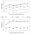
Pain ratings were collected subsequent to each of the 20s rectal balloon distensions. The mean pain ratings in response to each of the seven rectal distensions are shown for the group given the standard placebo instructions (top) and the enhanced placebo instructions (bottom). Pain ratings after intra-rectal lidocaine gel application are shown in the top panel.
Figure 2
Figure 2
Significant differences (FDR ≤ 0.05) between the standard placebo and rectal lidocaine conditions (i.e., s-PL > Lidocaine) identify areas wherein lidocaine was associated with a decrease in both pain and neural activity. This is a representative image of brain regions in which the magnitude of neural activity was significantly greater in the standard placebo compared to the rectal lidocaine condition. These areas are associated with afferent processing of pain and are in bold italics among the areas listed in Table 2.
Figure 3
Figure 3
Left image: significant differences (FDR ≤ 0.05) between the two placebo conditions in which the magnitude of activity was greater in the enhanced placebo condition than in the standard condition (e-PL > s-PL). This image presents representative brain regions that are among those listed in Table 3. Right: Representative BOLD activity curves showing greater activity in ePL than in s-PL (Right lentiform nucleus at top left, left and right superior temporal gyrus top right and lower left respectively). The lower right graph shows average activity from the 15 areas listed in Table 3. Many of these regions are typically associated with language-related functions, semantic processing, and memory. Note that in all cases the separation of the curves for the e-PL condition (solid line) and s-PL condition (dashed line) begins very near the stimulus onset and becomes increasing larger as the stimulus continues.
Figure 4
Figure 4
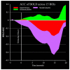
The graph shows average change in BOLD area under the curve (AUC), across time from the 15 areas listed in Table 3. Many of these regions are typically associated with language-related functions, semantic processing, and memory. Note the sharp decreases in area in the NH and standard placebo conditions compared to the enhanced placebo condition. AUC was calculated using the Trapezoid Rule (i.e., divided the time course into trapezoids, compute each area, sum the areas).
Figure 5
Figure 5
Left image: significant differences (FDR ≤ 0.05) between the two placebo conditions in which the magnitude of activity was greater in the standard placebo condition than in the enhanced placebo condition (s-PL > e-PL). This image presents representative brain regions, including those known to be involved in pain-related processing (thalamus, anterior cingulate cortex BA 32). Right: representative BOLD activity curves showing greater activity in the s-PL (dashed lines) than in the e-PL condition (solid lines), thereby indicating decreased pain-related activity in the latter condition. The left upper graph is based on data from the thalamus, the upper right graph on data from left anterior cingulate, and lower left graph on data from right posterior insula. The lower right graph shows average activity from the 21 areas listed in Table 4. Some of these regions are associated with pain processing as indicated by italicized bold regions listed in Table 4. Note that in all cases the separation of the curves for the e-PL condition (solid line) and s-PL condition (dashed line) begins very near the stimulus onset and becomes increasing larger as the stimulus continues.
Figure 6
Figure 6
Significant differences (FDR ≤ 0.05) between the enhanced placebo and rectal lidocaine conditions. This contrast identified representative brain regions wherein the e-PL condition was accompanied by increased activity in comparison to the lidocaine condition (see also Figure 1). Similar to regions shown in Figure 3, these regions reflect verbal/semantic processes, short and long term memory, and top-down processing in the e-PL condition as compared to the local anesthesia condition.

References

    1. Amanzio M, Benedetti F. Neuropharmacological dissection of placebo analgesia: expectation-activated opioid systems versus conditioning-activated specific subsystems. J Neurosci. 1999;19:484–494. - PMC - PubMed
    1. Atlas LY, Wager TD. How expectations shape pain. Neurosci Lett. 2012;520:140–148. - PubMed
    1. Bar M, Aminoff E, Mason M, Fenske M. The units of thought. Hippocampus. 2007;17:420–428. - PubMed
    1. Benedetti F, Amanzio M. Mechanisms of the placebo response. Pulmonary pharmacology & therapeutics. 2013 - PubMed
    1. Bjorkedal E, Flaten MA. Expectations of increased and decreased pain explain the effect of conditioned pain modulation in females. J Pain Res. 2012;5:289–300. - PMC - PubMed

Publication types