. 2014 Apr:69:403-16.
doi: 10.1016/j.freeradbiomed.2014.01.001. Epub 2014 Jan 8.

Chronic ethanol ingestion induces oxidative kidney injury through taurine-inhibitable inflammation

Affiliations

Chronic ethanol ingestion induces oxidative kidney injury through taurine-inhibitable inflammation

Calivarathan Latchoumycandane et al. Free Radic Biol Med. 2014 Apr.

Abstract

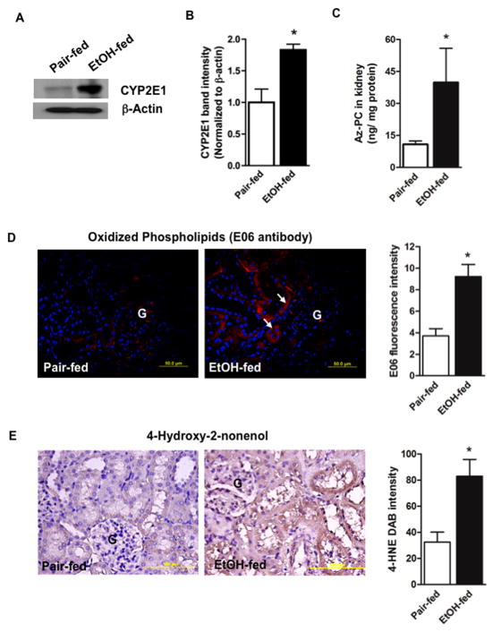
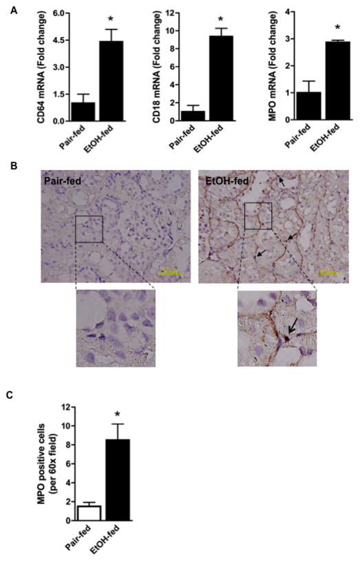
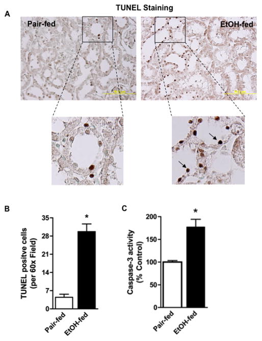
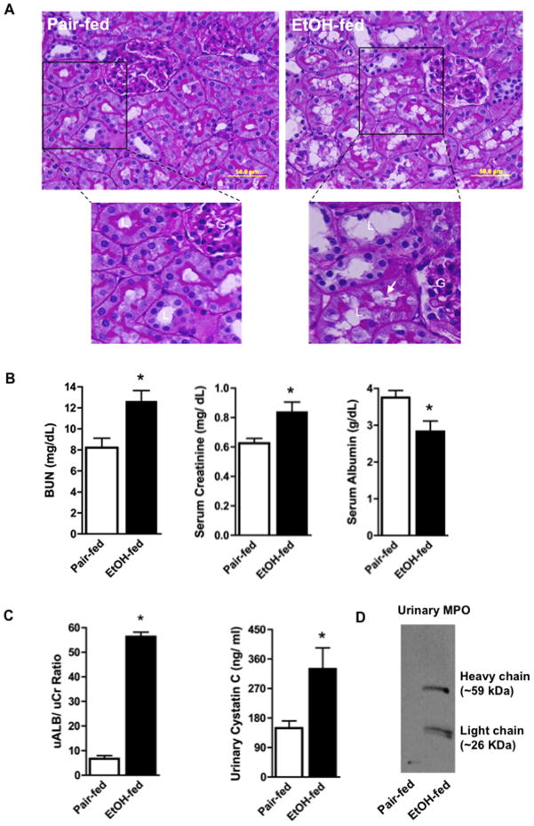
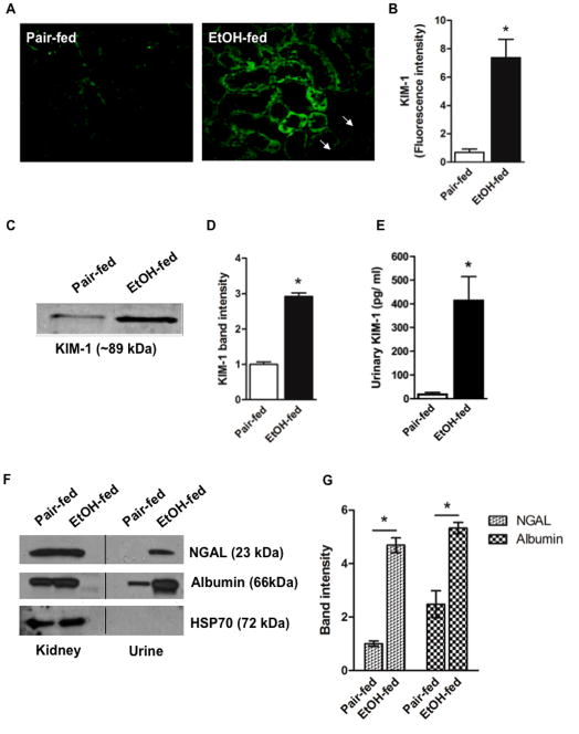
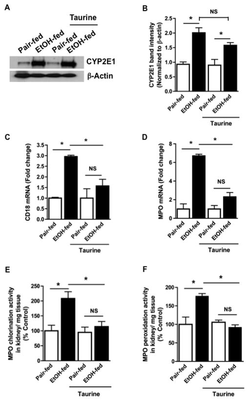
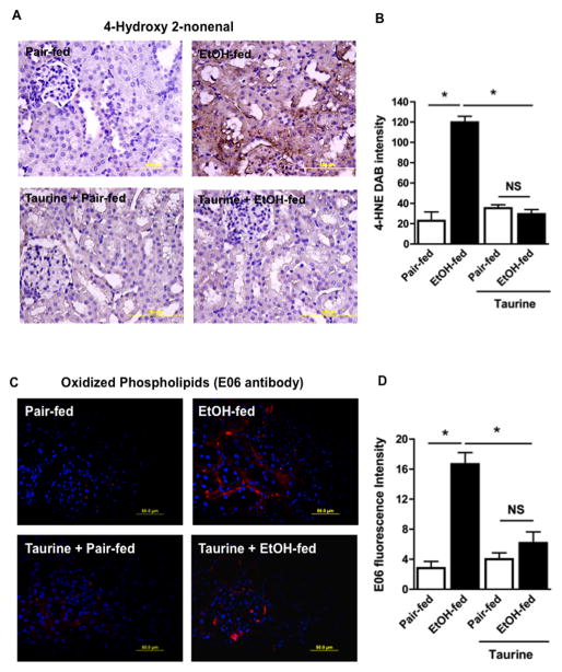
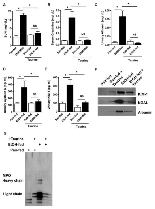
Chronic ethanol ingestion mildly damages liver through oxidative stress and lipid oxidation, which is ameliorated by dietary supplementation with the anti-inflammatory β-amino acid taurine. Kidney, like liver, expresses cytochrome P450 2E1 that catabolizes ethanol with free radical formation, and so also may be damaged by ethanol catabolism. Sudden loss of kidney function, and not liver disease itself, foreshadows mortality in patients with alcoholic hepatitis [J. Altamirano, Clin. Gastroenterol. Hepatol. 2012, 10:65]. We found that ethanol ingestion in the Lieber-deCarli rat model increased kidney lipid oxidation, 4-hydroxynonenal protein adduction, and oxidatively truncated phospholipids that attract and activate leukocytes. Chronic ethanol ingestion increased myeloperoxidase-expressing cells in kidney and induced an inflammatory cell infiltrate. Apoptotic terminal deoxynucleotidyl transferase nick-end labeling-positive cells and active caspase-3 increased in kidney after ethanol ingestion, with reduced filtration with increased circulating blood urea nitrogen (BUN) and creatinine. These events were accompanied by release of albumin, myeloperoxidase, and the acute kidney injury biomarkers kidney injury molecule-1 (KIM-1), neutrophil gelatinase-associated lipocalin, and cystatin c into urine. Taurine sequesters HOCl from myeloperoxidase of activated leukocytes, and taurine supplementation reduced renal lipid oxidation, reduced leukocyte infiltration, and reduced the increase in myeloperoxidase-positive cells during ethanol feeding. Taurine supplementation also normalized circulating BUN and creatinine levels and suppressed enhanced myeloperoxidase, albumin, KIM-1, and cystatin c in urine. Thus, chronic ethanol ingestion oxidatively damages kidney lipids and proteins, damages renal function, and induces acute kidney injury through an inflammatory cell infiltrate. The anti-inflammatory nutraceutical taurine effectively interrupts this ethanol-induced inflammatory cycle in kidney.

Keywords: Acute kidney injury; Free radicals; Inflammation; Kidney; Oxidized phospholipid; Reactive oxygen species; Taurine.


Figures

Figure 1
Figure 1. Chronic ethanol feeding induces oxidative stress in rat kidney
A) Kidney expresses ethanol-inducible CYP2E1. Western blotting shows CYP2E1 expression (55 kDa) in the crude homogenates (20 μg) of kidneys from pair-fed control and ethanol-fed rats. Equal loading of protein was confirmed by re-probing the blot with anti-β-actin antibody. B) Densitometry of CYP2E1 immunoblots. Band densities were determined with a Kodak Image Station and are expressed as mean ± SEM, n=3 with p<0.05 (*) considered significant. C) The oxidized phospholipid azelaoyl phosphatidylcholine increases in kidney after ethanol feeding. Quantitative mass spectrometry of palmitoyl azelaoyl phosphatidylcholine (Az-PC) using [2H]PAF as an internal standard was identified by elution time and the precursor/daughter ion transition m/z 667 → 184. n=6, p<0.05 (*). D) Oxidized phospholipid epitopes accumulate in the kidney of ethanol fed rats. Antigens were retrieved from formalin-fixed and paraffin-embedded kidney sections and stained for the E06 epitope of oxidized choline phospholipid adducts using Alexa Fluor568-conjugated secondary antibody (red fluorescence) with DAPI (blue) nuclear stain. n=3. “G” glomerulus; arrows, tubular wall red fluorescence. E06 immunofluorescence was quantified in 4 to 5 sections per kidney of pair-fed and ethanol-fed rats using ImageJ 1.47v software (NIH), and the data are expressed as mean ± SEM, n=3 p<0.05 (*). E) Reactive fatty acyl fragments adduct kidney protein after chronic ethanol feeding. Formalin-fixed and paraffin-embedded kidney sections were stained for the oxidative injury marker 4-hydroxynonenal (4-HNE) and developed using diaminobenzidine as the chromogen that generates a brown precipitate. The figure is representative of four to five sections from kidney of pair-fed and ethanol-fed rats (n=4) chronically ingested ethanol or pair-fed a control diet for 28 days. The 4-HNE DAB brown intensity was quantified in 4 to 5 sections per kidney using ImageJ and the data are expressed as mean ± SEM, n=4. p<0.05 (*).
Figure 2
Figure 2. Chronic ethanol feeding increases renal inflammation
A) Ethanol feeding increases inflammatory cell mRNA in kidney. Total RNA was extracted from kidney and mRNA was quantified by SYBR Green onestep reverse transcription-PCR for CD64 (left), CD18 (center), myeloperoxidase (MPO, right) and ribosomal S18. mRNA expression was normalized to S18 mRNA content and 2−ΔΔCT was used to calculate change. Data are expressed as mean ± SEM (CD64, n=10; CD18, n=10; MPO, n=4). p<0.05 (*) B) Myeloperoxidase positive cells increase in kidney during ethanol feeding. Sections of kidneys from rats fed the ethanol diet or their pair-fed controls were assayed for myeloperoxidase enzymatic activity. The reaction is marked by deposition of dark brown reaction product as described in “Methods”. These images are representative of four to five sections per kidney of pair-fed and ethanol-fed rats (n=4). The 60X inset shows granular myeloperoxidase activity stain associated with tubular walls and an infiltrating cell (arrow). C) Quantification of myeloperoxidase positive cells. Myeloperoxidase positive cells were enumerated in 3 random kidney sections at 60X with the data expressed as mean ± SEM (n=4). p<0.05 (*)
Figure 3
Figure 3. Chronic ethanol feeding induces renal tubular apoptosis
A) Chronic ethanol ingestion increases kidney cell death. Kidneys of control and ethanol-fed rats were harvested, fixed, sectioned, and stained for TUNEL positive cells using a commercial kit that develops a dark brown TUNEL reaction product. The 60X inset shows these cells primarily were tubular. B) Ethanol feeding significantly increases TUNEL positive kidney cells. TUNEL positive cells were enumerated in 3 random kidney sections at 60X with the data expressed as mean ± SEM (n=4). p<0.05 (*) C) Ethanol feeding increases renal caspase-3 activity. Caspase-3 activity was determined in crude kidney homogenates of control- or ethanol-fed rats as described in “Methods”. Caspase-3 activity is given as a percent of control and data are expressed as mean ± SEM (n=6). p<0.05 (*)
Figure 4
Figure 4. Ethanol alters renal architecture and induces renal injury
A) Periodate-Schiff staining of kidneys from pair-fed control and ethanol-fed rats. Paraffin embedded kidney sections were deparaffinized and hydrated before being stained by periodic-acid Schiff/hematoxylin. “G”, glomerulus; “L” lumen; arrow, loss of PAS positive microvilli. B) Ethanol feeding induces filtration defects. Blood urea nitrogen (BUN), creatinine, and albumin from rats pair-fed a control diet and ethanol-fed rats were measured as described in “Methods”. Data are expressed as mean ± SEM (n=8). p<0.05 (*) C) Ethanol feeding induces albuminuria and increases urinary cystatin c. Urinary albumin, creatinine, and cystatin c were quantified by commercial kits. Data are expressed as mean ± SEM (n=4). p≤0.05 (*) D) Proteolytically processed myeloperoxidase is shed into urine of ethanol fed rats. Western blot of myeloperoxidase of urine from control and ethanol-fed rats. The lanes contained equal volumes of urine since loading controls are not appropriate.
Figure 5
Figure 5. Chronic ethanol feeding induces AKI
A) Ethanol feeding induces renal KIM-1 expression. Kidney sections from pair-fed control rats and those fed ethanol were immunostained for KIM-1 and detected with Alexa Fluor488-conjugated secondary antibody. These immunofluorescent images are representative of four to five sections per kidney of pair-fed and ethanol-fed rats (n=4). B) Quantification of KIM-1 fluorescence. Image intensity was quantified using ImageJ, and expressed as mean ± SEM, n=4. p<0.05 (*) C) Urinary KIM-1 increases in response to ethanol ingestion. Western blot of KIM-1 in urine from pair-fed and ethanol-fed rats migrating at approximately 89 kDa. D) Quantification of urinary KIM-1 immunoblots. Relative band density was quantified in a Kodak Image Station, and the data expressed as mean ± SEM (n=3). p<0.05 (*) E) Immunoreactive KIM-1 increases in urine after ethanol feeding. Urinary KIM-1 was quantified by ELISA with the data expressed as mean ± SEM (n=8). p<0.05 (*) F) NGAL and albumin are shed from the kidneys of rats ingesting ethanol. Western blots of crude kidney homogenates or equal volumes of urine collected from ethanol-fed rats or their pair-fed controls were immunoblotted for NGAL, albumin, or cellular HSP70. The lack of HSP70 shows effective clearance of cellular debris from urine samples. G) Quantification of urinary AKI markers. Relative band intensities for NGAL and albumin were quantified in a Kodak Image Station, with data expressed as mean ± SEM (n=3). p<0.05 (*)
Figure 6
Figure 6. Dietary taurine ameliorates ethanol-induced renal inflammation and kidney injury
A) Taurine does not alter CYP2E1 expression in kidney of ethanol-fed rats. Western blotting of CYP2E1 expression (55 kDa) in crude homogenates of kidneys from rats ingesting ethanol and/or taurine as shown. Equal loading of protein was confirmed by re-probing the blot with anti-β-actin antibody. B) Quantitation of CYP2E1 immunoblot band density. Densitometry for CYP2E1 accumulated as stated are expressed as mean SEM, n=3, determined by two-way analysis of variance. p<0.05 (*) C) Taurine suppresses infiltration of CD64-expressing cells into kidney of ethanol-fed rats. Taurine was included, or not, in the liquid diet fed with or without ethanol for 28 days. mRNA for CD18 in kidney homogenates was assessed as in panel 2A with data expressed as mean ± SEM (n=4) by two way ANOVA. p<0.05 (*) D) Taurine suppresses infiltration of CD18-expressing cells into kidney of ethanol-fed rats. Taurine and/or ethanol ingestion were as stated in the panel before renal myeloperoxidase mRNA in kidney homogenate was assessed as in panel 2A. The data are expressed as mean ± SEM (n=4) analyzed by two factor analysis of variance. p<0.05 (*) Dietary taurine supplementation abolishes the ethanol-induced increase in kidney myeloperoxidase activity. Chlorination (E) and peroxidation (F) activities were separately determined in the crude homogenates of rat kidney from ethanol- or control-fed rats additionally, or not, ingesting taurine. These myeloperoxidase half reactions were measured as described in “Methods’ with enzymatic activity expressed as a percent of control. The data are the mean ± SEM (n=4) by two-way ANOVA. p<0.05 (*)
Figure 7
Figure 7. Taurine supplementation ameliorates ethanol-induced renal oxidative injury
A) Dietary taurine suppresses 4-hydroxynonenal protein modification induced by ethanol ingestion. Kidneys from rats fed as described in the panels were harvested, sectioned, fixed, and immunostained with anti-4-hydroxynonenal (4-HNE) antibody as in Fig. 1E. Panels are representative of four sections from each of four animals. B. 4-Hydroyxnonenal image quantitation. DAB staining was quantified by ImageJ in 3 random kidney sections at 60X with the data expressed as mean ± SEM using two-way ANOVA (n=4). p<0.05 (*) C) Dietary taurine suppresses the ethanol-induced increase in renal choline phospholipid oxidation adducts. Kidneys of rats fed as described were harvested, sectioned, fixed, and immunostaining with E06 antibody as in Fig. 1D. Panels are representative of four sections from each of four animals. D. E06 image quantitation. E06 fluorescence was quantified by ImageJ in 3 random kidney sections at 60X with the data expressed as mean ± SEM by two-way analysis of variance (n=4). p<0.05 (*)
Figure 8
Figure 8. Taurine supplementation abolishes ethanol-induced renal dysfunction and development of AKI
Dietary taurine abolishes renal filtration defects induced by chronic ethanol ingestion. Sera of rats fed ethanol or pair-fed a control diet, received dietary taurine, or not, was assayed for A) BUN or B) creatinine as in Fig. 4B. The data are analyzed by two factor analysis of variance and shown as mean ± SEM (n=4). p<0.05 (*) C) Ethanol-induced release of albumin, D) cystatin C, and E) KIM-1 to urine was blocked by dietary taurine. Urinary markers were quantitated as in Fig. 4C and expressed as mean ± SEM (n=4) and analyzed by two-way ANOVA. p<0.05 (*) F) Taurine supplementation suppresses protein shedding to urine. Western blots of urinary KIM-1, NGAL, and albumin, using antibodies distinct from those of the preceding panel, were detected as described in Fig. 5F. G) Taurine supplementation ameliorates ethanol-induced myeloperoxidase shedding to urine. Myeloperoxidase fragments were detected in equal volumes of urine by western blotting with anti-myeloperoxidase as in Fig. 4D.

References

    1. O’Shea RS, Dasarathy S, McCullough AJ. Alcoholic liver disease. Hepatology. 2010;51:307–328. - PubMed
    1. Lucey MR, Mathurin P, Morgan TR. Alcoholic hepatitis. New Engl’d J Med. 2009;360:2758–2769. - PubMed
    1. Dey A, Cederbaum AI. Alcohol and oxidative liver injury. Hepatology. 2006;43:S63–74. - PubMed
    1. Lu Y, Wu D, Wang X, Ward SC, Cederbaum AI. Chronic alcohol-induced liver injury and oxidant stress are decreased in cytochrome P4502E1 knockout mice and restored in humanized cytochrome P4502E1 knock-in mice. Free Radic Biol Med. 2010;49:1406–1416. - PMC - PubMed
    1. Kono H, Rusyn I, Uesugi T, Yamashina S, Connor HD, Dikalova A, Mason RP, Thurman RG. Diphenyleneiodonium sulfate, an NADPH oxidase inhibitor, prevents early alcohol-induced liver injury in the rat. Am J Physiol Gastrointest Liver Physiol. 2001;280:G1005–1012. - PubMed

Publication types

LinkOut - more resources