. 2014 Feb;21(2):175-179.
doi: 10.1038/nsmb.2753. Epub 2014 Jan 12.

RNA polymerase II termination involves C-terminal-domain tyrosine dephosphorylation by CPF subunit Glc7

Affiliations

RNA polymerase II termination involves C-terminal-domain tyrosine dephosphorylation by CPF subunit Glc7

Amelie Schreieck et al. Nat Struct Mol Biol. 2014 Feb.

Abstract

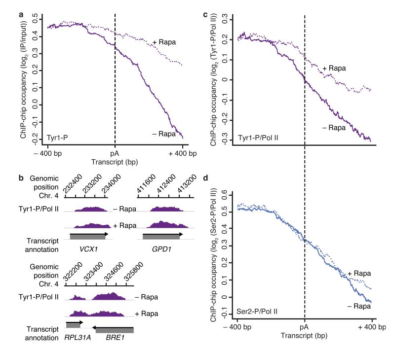
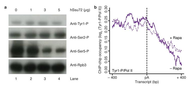
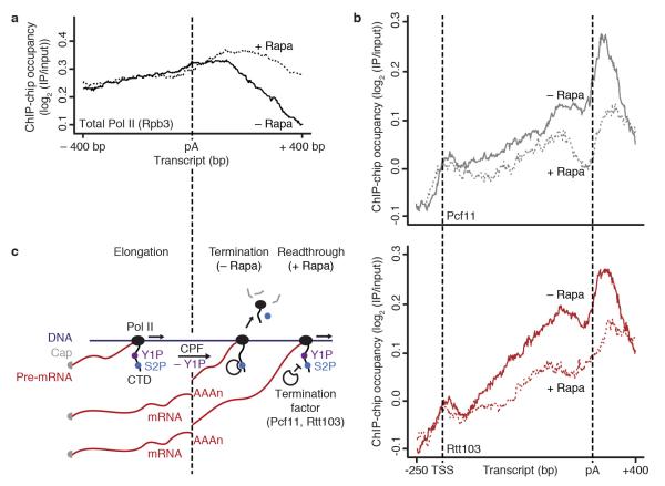
At the 3' ends of protein-coding genes, RNA polymerase (Pol) II is dephosphorylated at tyrosine residues (Tyr1) of its C-terminal domain (CTD). In addition, the associated cleavage-and-polyadenylation factor (CPF) cleaves the transcript and adds a poly(a) tail. Whether these events are coordinated and how they lead to transcription termination remains poorly understood. Here we show that CPF from Saccharomyces cerevisiae is a Pol II-CTD phosphatase and that the CPF subunit Glc7 dephosphorylates Tyr1 in vitro. In vivo, the activity of Glc7 is required for normal Tyr1 dephosphorylation at the polyadenylation site, for recruitment of termination factors Pcf11 and Rtt103 and for normal Pol II termination. These results show that transcription termination involves Tyr1 dephosphorylation of the CTD and indicate that pre-mRNA processing by CPF and transcription termination are coupled via Glc7-dependent Pol II-Tyr1 dephosphorylation.


Figures

Figure 1
Figure 1
CPF subunit Glc7 is a Pol II CTD Tyr1 phosphatase in vitro. (a) SDS-PAGE analysis of purified yeast CPF. An asterisk marks the tagged protein Ref2; the phosphatases Ssu72 and Glc7 are underlined. Identities of bands confirmed by mass-spectrometry are labeled. (b) In vitro dephosphorylation assay measuring CPF activity towards phosphorylated Pol II CTD as shown by Western blotting with antibodies against Pol II subunit Rpb3 and Tyr1-phosphorylated (Tyr1-P), Ser2-P and Ser5-P CTD residues (1Y26, 3D12, 3E10 and 3E8 antibodies, respectively). For lanes 6–13, 10 mM EDTA or 200 nM microcystin were included in the reactions. Uncropped versions of blots can be found in Supplementary Fig. 4a.
Figure 2
Figure 2
Glc7 is required for Tyr1 but not Ser2 dephosphorylation in vivo. (a) Metagene analysis for genome-wide ChIP occupancy of Tyr1-phosphorylated Pol II around polyA (pA) sites in the Glc7 anchor-away strain, treated with rapamycin to deplete Glc7 from the nucleus (+ Rapa, violet dotted line) or untreated (− Rapa, violet solid line). Averaged ChIP-chip signals are plotted as the median signal (log2 (IP/input)) at each genomic position over a set of 619 representative genes (Online Methods), normalized to have approximately equal occupancy levels upstream (around −400 bp) of the pA site (dashed black line). The profiles are shown from 400 nucleotides upstream to 400 nucleotides downstream of the pA site. Non-normalized data averaged over the entire gene length are shown in Supplementary Fig. 2. Experiments were performed in biological duplicates. (b) Profiles of Tyr1-P levels normalized to total Pol II (Fig. 4a) on selected genes, smoothed by a 150 nt window running median are shown in purple. Below, grey boxes indicate transcripts on the Watson (top) and Crick strands (bottom). (c,d) ChIP-chip occupancy profiles of Tyr1- (c) and Ser2- (d) phosphorylated Pol II over 619 genes aligned at the pA site (dashed line) and normalized against the corresponding Rpb3-profile (Fig. 4a) without and with rapamycin (solid and dotted lines, respectively). Experiments were performed in biological duplicates.
Figure 3
Figure 3
Ssu72 does not change Tyr1 phosphorylation levels in vitro and in vivo. (a) In vitro dephosphorylation assay of Pol II CTD with recombinant human Ssu72, monitored by Western blotting with antibodies against Pol II subunit Rpb3 and Tyr1-phosphorylated (Tyr1-P), Ser2-P and Ser5-P CTD residues (1Y26, 3D12, 3E10 and 3E8 antibodies, respectively). Uncropped versions of blots can be found in Supplementary Fig. 4b. (b) ChIP-chip occupancy profiling of Tyr1-phosphorylated Pol II over 619 genes aligned at the pA site (dashed line) and normalized against the corresponding Rpb3 profile without and with rapamycin treatment (solid and dotted lines, respectively) in a Ssu72 anchor away strain. Profiles in a region from 400 nucleotides upstream to 400 nucleotides downstream of the pA site are shown. This experiment was performed in biological duplicate.
Figure 4
Figure 4
Tyr1 dephosphorylation by Glc7 is required for normal termination factor recruitment and transcription termination in vivo. (a) Metagene analysis for genome-wide ChIP occupancy of total Pol II (subunit Rpb3) around polyA (pA) sites in the Glc7 anchor-away strain with rapamycin treatment (+ Rapa, black dotted line) and untreated (− Rapa, black line). Averaged ChIP-chip signals are shown as the median signal (log2 (IP/input)) at each genomic position over a set of 619 representative genes (Online Methods) and normalized to have approximately equal occupancy levels upstream (around −400 bp) of the pA site. (b) ChIP-chip occupancy profiling of TAP-tagged Pcf11 (top) and Rtt103 (bottom) in the Glc7 anchor-away strain, after treatment with rapamycin (+ Rapa, dotted lines) and untreated (− Rapa, solid lines). The profiles show ChIP-chip signals averaged by taking the median signal (log2 (IP/input)) at each genomic position over a set of representative medium length genes (1238 ± 300 bp, n = 339, Online Methods). TSS, transcription start site. Experiments in (a,b) were performed in biological duplicates. (c) Model for the Pol II elongation-termination transition. DNA is dark blue, RNA is dark red, Pol II and its CTD are black; Ser2-P is blue and Tyr1-P is purple. For details see within text.

References

    1. Brannan K, Bentley DL. Control of Transcriptional Elongation by RNA Polymerase II: A Retrospective. Genet Res Int. 2012;2012:170–173. - PMC - PubMed
    1. Buratowski S. Progression through the RNA polymerase II CTD cycle. Mol. Cell. 2009;36:541–546. - PMC - PubMed
    1. Corden JL. Transcription. Seven ups the code. Science. 2007;318:1735–1736. - PubMed
    1. Jeronimo C, Bataille AR, Robert F. The Writers, Readers, and Functions of the RNA Polymerase II C-Terminal Domain Code. Chem. Rev. 2013;113:8491–8522. - PubMed
    1. Komarnitsky P, Cho EJ, Buratowski S. Different phosphorylated forms of RNA polymerase II and associated mRNA processing factors during transcription. Genes Dev. 2000;14:2452–2460. - PMC - PubMed

METHODS REFERENCES

    1. Passmore LA, et al. Doc1 mediates the activity of the anaphase-promoting complex by contributing to substrate recognition. EMBO J. 2003;22:786–796. - PMC - PubMed
    1. Sydow JF, et al. Structural basis of transcription: mismatch-specific fidelity mechanisms and paused RNA polymerase II with frayed RNA. Mol. Cell. 2009;34:710–721. - PubMed
    1. Chapman RD, et al. Transcribing RNA polymerase II is phosphorylated at CTD residue serine-7. Science. 2007;318:1780–1782. - PubMed
    1. Mayer A, et al. The spt5 C-terminal region recruits yeast 3′ RNA cleavage factor I. Mol. Cell. Biol. 2012;32:1321–1331. - PMC - PubMed
    1. Nagalakshmi U, et al. The transcriptional landscape of the yeast genome defined by RNA sequencing. Science. 2008;320:1344–1349. - PMC - PubMed

Publication types

MeSH terms