. 2014 Mar;6(3):347-57.
doi: 10.1002/emmm.201303133. Epub 2014 Jan 10.

Heteropathogenic virulence and phylogeny reveal phased pathogenic metamorphosis in Escherichia coli O2:H6

Affiliations

Heteropathogenic virulence and phylogeny reveal phased pathogenic metamorphosis in Escherichia coli O2:H6

Martina Bielaszewska et al. EMBO Mol Med. 2014 Mar.

Abstract

Extraintestinal pathogenic and intestinal pathogenic (diarrheagenic) Escherichia coli differ phylogenetically and by virulence profiles. Classic theory teaches simple linear descent in this species, where non-pathogens acquire virulence traits and emerge as pathogens. However, diarrheagenic Shiga toxin-producing E. coli (STEC) O2:H6 not only possess and express virulence factors associated with diarrheagenic and uropathogenic E. coli but also cause diarrhea and urinary tract infections. These organisms are phylogenetically positioned between members of an intestinal pathogenic group (STEC) and extraintestinal pathogenic E. coli. STEC O2:H6 is, therefore, a 'heteropathogen,' and the first such hybrid virulent E. coli identified. The phylogeny of these E. coli and the repertoire of virulence traits they possess compel consideration of an alternate view of pathogen emergence, whereby one pathogroup of E. coli undergoes phased metamorphosis into another. By understanding the evolutionary mechanisms of bacterial pathogens, rational strategies for counteracting their detrimental effects on humans can be developed.


Figures
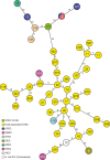
Figure 1
Figure 1
Phylogenetic relationships of STEC O2:H6 to intestinal and extraintestinal pathogenic Escherichia coli and to E. coli. K-12. Minimum spanning tree based on MLST and rMLST allelic profiles portraying the clonal relationships of STEC O2:H6 to HUS-associated STEC (HUSEC collection) (Mellmann et al, 2008), other intestinal pathogenic E. coli (EPEC, ETEC, EIEC, EAEC, AIEC), prototypic ExPEC including UPEC and MNEC isolates, and a non-pathogenic E. coli strain K-12 (MG1655). Isolates are described in supplementary Table S1. Each circle represents a given allelic profile (combination of MLST and rMLST loci) and is named with the MLST sequence type. The different groups of strains are distinguished by colors of the circles. The numbers on the connecting lines illustrate the number of differing alleles.
Figure 2
Figure 2
Phylogenetic relationships of STEC O2:H6 to prototypic UPEC, AIEC and most closely related and prototypic HUS-associated STEC based on whole genome sequencing. Minimum spanning tree is based on allelic profiles of 2827 genes present in all strains investigated (see supplementary Table S1). The different pathotypes are distinguished by colors of the circles and the serotypes and strain numbers (in parentheses) are given.
Figure 3
Figure 3
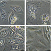
Production of colibactin by STEC O2:H6. HeLa cells were cocultured with bacteria (4 h), washed and incubated in gentamicin-supplemented medium (48 h). The DNA content was determined by flow cytometry and morphological changes were assessed microscopically. Bar = 100 μm. A Uninfected (control) cells were mostly in the G1 phase of the cell cycle (2n DNA) and retained normal morphology. B–C Cells infected with clb-positive STEC O2:H6 strain 05-00787 (B) or the prototypic clb-harboring strain IHE3034 (C) were arrested in the G2 phase (4n DNA) and converted into megalocytes. (The phenotype shown in (B) was produced by each of three clb-positive, α-hlyA-negative and cnf1-negative STEC O2:H6). D Cells infected with clb-negative Stx2-producing O2:H27 isolate displayed neither G2 arrest nor distension.
Figure 4
Figure 4
Contact-dependent growth inhibition mediated by STEC O2:H6. Ampicillin-resistant target strain MG1655/pBlueskript KS II(+) was cultured alone or in mixture with log-phase culture of each inhibitor (inhibitor-to-target ratio 50:1) including cdiAB-positive STEC O2:H6 strains 03-08304, 05-06793, 04-03909, prototypic cdiAB-harboring strain EC93, or cdiAB-negative STEC O2:H27. At each indicated time point, the growth of the target strain (CFU/ml) was determined by plating 10-fold culture dilutions on LB agar with ampicillin. Data represent means ± standard deviations of three independent experiments. *< 0.01 (unpaired Student′s t-test), differences between growth of the target strain alone and in coculture with each respective inhibitor.
Figure 5
Figure 5
Vacuolization induced by STEC O2:H6. CHO cells were exposed to sterile culture supernatants of tested strains and presence of vacuoles was sought microscopically after 24 h. Bar = 20 μm. A–B vat-positive STEC O2:H6 strains 05-00787 (A) and 05-06739 (B). (Vacuolization similar to that displayed by these two strains was elicited by all STEC O2:H6 isolates). C vat-containing UPEC strain J96 (positive control). D Uninfected cells (negative control).
Figure 6
Figure 6
Urovirulence of STEC O2:H6 strains. Bladder (A) and kidney (B) colonization levels were determined 72 h after transurethral inoculation of mice with UPEC strain 536 (positive control), the STEC O2:H6 strains 05-00787, 04-00955, and 03-08304, or non-pathogenic Escherichia coli K-12 strain MG1655 (negative control). Horizontal bars represent the mean CFU number of each strain per gram of tissue; the whiskers display the respective standard error of the mean. Significant differences in the bacterial organ load compared to the negative control are indicated by asterisks.

References

    1. Achtman M, Mercer A, Kusecek B, Pohl A, Heuzenroeder M, Aaronson W, Sutton A, Silver RP. Six widespread bacterial clones among Escherichia coli K1 isolates. Infect Immun. 1983;39:315–335. - PMC - PubMed
    1. Aoki SK, Pamma R, Hernday AD, Bickham JE, Braaten BA, Low DA. Contact-dependent inhibition of growth in Escherichia coli. Science. 2005;309:1245–1248. - PubMed
    1. Bielaszewska M, Fell M, Greune L, Prager R, Fruth A, Tschäpe H, Schmidt MA, Karch H. Characterization of cytolethal distending toxin genes and expression in Shiga toxin-producing Escherichia coli strains of non-O157 serogroups. Infect Immun. 2004;72:1812–1816. - PMC - PubMed
    1. Bielaszewska M, Friedrich AW, Aldick T, Schurk-Bulgrin R, Karch H. Shiga toxin activatable by intestinal mucus in Escherichia coli isolated from humans: predictor for a severe clinical outcome. Clin Infect Dis. 2006;43:1160–1167. - PubMed
    1. Bielaszewska M, Mellmann A, Bletz S, Zhang W, Kock R, Kossow A, Prager R, Fruth A, Orth-Holler D, Marejkova M, et al. Enterohemorrhagic Escherichia coli O26:H11/H−: a new virulent clone emerges in Europe. Clin Infect Dis. 2013;56:1373–1381. - PubMed

Publication types

MeSH terms

Associated data