. 2014 Feb;46(2):166-70.
doi: 10.1038/ng.2873. Epub 2014 Jan 12.

Recurrent mutations in epigenetic regulators, RHOA and FYN kinase in peripheral T cell lymphomas

Affiliations

Recurrent mutations in epigenetic regulators, RHOA and FYN kinase in peripheral T cell lymphomas

Teresa Palomero et al. Nat Genet. 2014 Feb.

Abstract

Peripheral T cell lymphomas (PTCLs) are a heterogeneous and poorly understood group of non-Hodgkin lymphomas. Here we combined whole-exome sequencing of 12 tumor-normal DNA pairs, RNA sequencing analysis and targeted deep sequencing to identify new genetic alterations in PTCL transformation. These analyses identified highly recurrent epigenetic factor mutations in TET2, DNMT3A and IDH2 as well as a new highly prevalent RHOA mutation encoding a p.Gly17Val alteration present in 22 of 35 (67%) angioimmunoblastic T cell lymphoma (AITL) samples and in 8 of 44 (18%) PTCL, not otherwise specified (PTCL-NOS) samples. Mechanistically, the RHOA Gly17Val protein interferes with RHOA signaling in biochemical and cellular assays, an effect potentially mediated by the sequestration of activated guanine-exchange factor (GEF) proteins. In addition, we describe new and recurrent, albeit less frequent, genetic defects including mutations in FYN, ATM, B2M and CD58 implicating SRC signaling, impaired DNA damage response and escape from immune surveillance mechanisms in the pathogenesis of PTCL.


Figures

Figure 1
Figure 1. RHOA mutations in PTCLs
(a) Schematic representation of the structure of the RHOA protein. RHOA mutations identified by targeted amplicon resequencing in PTCL samples are shown (n=64). Multiple circles in the same amino acid position account for multiple patients with the same variant. (b) Differential distribution of RHOA mutations in all PTCL categories, PTCL NOS and AITLs. (c) Distribution of RHOA p.Gly17Val, TET2, DNMT3A and IDH2 mutations in major PTCL groups (AITL, n=30; PTCL NOS, n=17; ALCL ALK+, n=4; and ALCL ALKL–, n=2). Colored boxes indicate the presence of mutations in the indicated genes (rows) in each patient sample (columns).
Figure 2
Figure 2. Functional characterization of the RHOA Gly17Val protein
(a) GFP fluorescence micrographs of HEK293T cells expressing GFP, GFP-RHOA, constitutively active GFP-RHOA Q63L, dominant negative GFP-RHOA Thr19Asn and GFP-RHOA Gly17Val protein. Scale bar = 10μm. (b) Immunofluorescence analysis of stress fiber formation in HeLa cells expressing GFP, GFP-RHOA, GFP-RHOA Gln63Leu, GFP-RHOA Thr19Asn and GFP-RHOA Gly17Val protein shown in green. Actin fibers stained with phalloidin are shown in red and cell nuclei stained with DAPI are shown in blue. Scale bar = 20μm. (c) Western blot analysis of GTP-bound HA-RHOA in rhotekin pull downs from Jurkat cells expressing wild type HA-RHOA, constitutively active HARHOA Gln63Leu, dominant negative HA-RHOA Thr19Asn and the PTCL associated HA-RHOA Gly17Val protein. (d) Fluorescence polarization analysis of mant-GTP loading to GST-RHOA, GST-RHOA Gly17Ala and GST-RHOA Gly17Val in response to MCF2L/DBS stimulation. (c) Western blot analysis of ARHGEF1 GEF protein pulled down with GST-RHOA, GST-RHOA Gly17Ala and GST-RHOA Gly17Val from Jurkat cell lysates in basal conditions and upon serum (FBS) stimulation. Pounceau S staining of bait protein loading is shown at the bottom. Representative images from at least two independent experiments are shown in (a) and (b). Data in (d) shows average ± s.d. from triplicate samples.
Figure 3
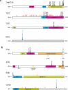
Figure 3. DNMT3A, TET2, IDH2, FYN, ATM, TET3, B2M and CD58 mutations in PTCLs
a) Schematic representation of DNMT3A, TET2, TET3 and IDH2 proteins showing DNA methylation and hydroxymethylation related mutations in PTCL patients via exome sequencing (n=12) and amplicon resequencing (n=64). b) Schematic representation of FYN, ATM, B2M and CD58 protein variants identified in PTCL samples (n=12) and amplicon resequencing (n=64). Solid circles indicate predicted amino acid substitutions. The position of truncating mutations is indicated with red open circles. Multiple circles in the same amino acid position account for multiple patients with the same variant.
Figure 4
Figure 4. Structure modeling and functional characterization of FYN mutations identified in PTCLs
(a) Analysis of FYN activation via phospho-SRC immunoblotting in Rat1A cells infected wild type and PTCL associated FYN mutants expressing retroviruses. (b) Analysis of FYN activation via phosphor-SRC immunoblotting of FYN immunoprecipitates from Rat1A cells infected with wild type and PTCL associated FYN mutants expressing retroviruses. (c) Molecular ribbon representation of wild type FYN protein structure showing the positioning of the FYN SH2 domain and the C terminal Tyr531 phosphosite. (d) Structure modeling of FYN Tyr531His, FYN Arg176Cys and FYN Leu174arg mutant proteins. (e) Analysis of wild type GSTSH2-FYN interaction with C-terminal FYN peptides corresponding to wild type Tyr531 FYN, wild type P-Tyr531 FYN and mutant Tyr531His FYN via Western blot analysis of GST-SH2-FYN proteins in streptavidin-biotin C-terminal FYN peptide pull downs. Experiment was replicated twice. (f) Analysis of P-Tyr531 FYN C-terminal FYN peptide interaction with wild type GST-SH2-FYN and GST-SH2-FYN Leu174Arg and GST-SH2-FYN Arg176Cys mutant proteins via Western blot analysis of GST-SH2-FYN proteins in streptavidin-biotin P-Tyr531 C-terminal FYN peptide pull downs. (g) Western blot analysis of CSK inhibition of FYN activity in HeLa cells expressing wild type and PTCL associated FYN mutant proteins. (h) Western blot analysis of dasatinib inhibition of FYN activity in HEK293T cells expressing PTCL associated FYN mutant proteins. (i, j) Analysis of dasatinib effects on FYN phosphorylation (i) and relative cell growth (j) in transformed Rat1A cells expressing the constitutively active FYN Tyr531His or the dasatinib-resistant FYN Thr342Ile Tyr531His double mutant protein. Data in (j) shows average ± s.d. from triplicate samples. P values were calculated using the two-tailed Student's t test.

References

    1. Armitage JO. The aggressive peripheral T-cell lymphomas: 2012 update on diagnosis, risk stratification, and management. Am J Hematol. 2012;87:511–9. - PubMed
    1. Rudiger T, et al. Peripheral T-cell lymphoma (excluding anaplastic large-cell lymphoma): results from the Non-Hodgkin's Lymphoma Classification Project. Ann Oncol. 2002;13:140–9. - PubMed
    1. Schiller MR. Coupling receptor tyrosine kinases to Rho GTPases--GEFs what's the link. Cell Signal. 2006;18:1834–43. - PubMed
    1. Bar-Sagi D, Hall A. Ras and Rho GTPases: a family reunion. Cell. 2000;103:227–38. - PubMed
    1. Vega FM, Ridley AJ. Rho GTPases in cancer cell biology. FEBS Lett. 2008;582:2093–101. - PubMed

Publication types

MeSH terms