Review
. 2014 Apr;466(4):661-75.
doi: 10.1007/s00424-013-1434-6. Epub 2014 Jan 11.

T-type channels buddy up

Affiliations
Review

T-type channels buddy up

Ray W Turner et al. Pflugers Arch. 2014 Apr.

Abstract

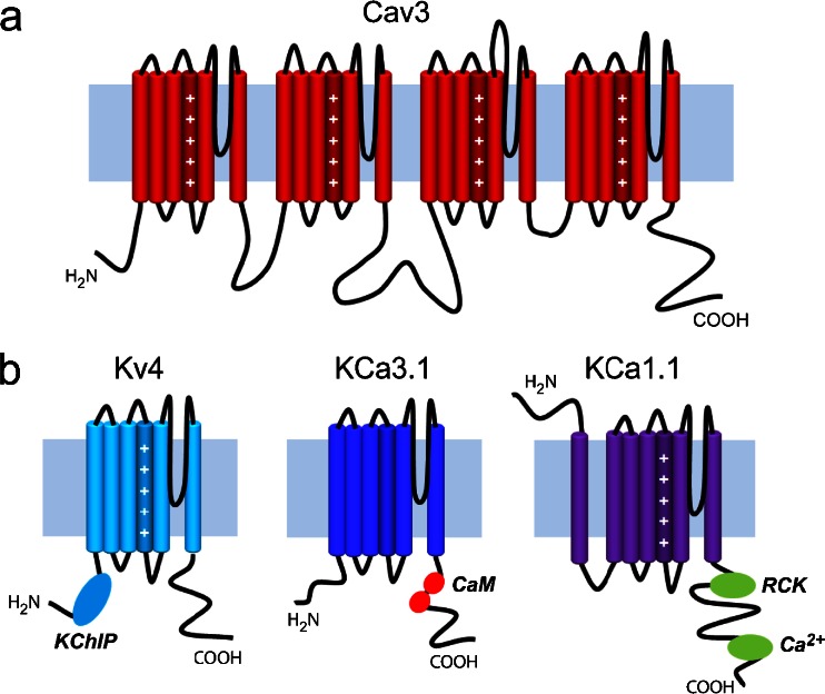
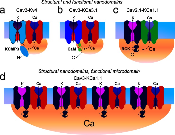
The electrical output of neurons relies critically on voltage- and calcium-gated ion channels. The traditional view of ion channels is that they operate independently of each other in the plasma membrane in a manner that could be predicted according to biophysical characteristics of the isolated current. However, there is increasing evidence that channels interact with each other not just functionally but also physically. This is exemplified in the case of Cav3 T-type calcium channels, where new work indicates the ability to form signaling complexes with different types of calcium-gated and even voltage-gated potassium channels. The formation of a Cav3-K complex provides the calcium source required to activate KCa1.1 or KCa3.1 channels and, furthermore, to bestow a calcium-dependent regulation of Kv4 channels via associated KChIP proteins. Here, we review these interactions and discuss their significance in the context of neuronal firing properties.


Figures

Fig. 1
Fig. 1
Transmembrane topology of the Cav3 α1 subunit and the three types of potassium channel subunits discussed in this review. a Cav3 channels are comprised of four homologous transmembrane domains that are connected via large cytoplasmic linkers. Each of these domains contains six-membrane spanning helices plus a re-entrant pore loop. Voltage sensors are formed by positive charges on the fourth transmembrane helix in each domain. b Potassium channel subtypes recognized to form ion signaling complexes with Cav3 calcium channels. Kv4 channels are similar to a single domain of Cav3 channels, and four of these combine to form a functional channel. KChIP molecules attached to the N-terminus region act as calcium sensors. KCa3.1 channels are similar in structure to Kv4 channels but lack voltage sensors. They are purely activated by calcium via interactions with calmodulin (CaM) on the channel C-terminus. Finally, KCa1.1 channels show a slightly different membrane topology, with an additional transmembrane helix that places the N-terminus at the extracellular end of the protein. Calcium is sensed by RCK domains and a calcium bowl located on the C-terminus region of the channel
Fig. 2
Fig. 2
Cav3 calcium channels associate with KCa3.1 channels to regulate temporal summation of EPSPs. ac Dual-label immunocytochemistry for Cav3.2 (a) and KCa3.1 (b) reveals protein colocalized (arrows) at the soma (asterisks) and restricted segments of dendrites (c). d RT-PCR reveals KCa3.1 and MRF-1 mRNA in the whole cerebellum (left) and KCa1.1, KCa2.2, and KCa3.1 but not MRF-1 in single Purkinje cell cytoplasmic extracts (right). e KCa3.1 protein coimmunoprecipitates with Cav3.2 from rat cerebellar lysate. f On-cell channel recordings (+30-mV pipette potential) before and after perfusing the calcium ionophore A23187 (2 μM) and blocked by TRAM-34 (100 nM). Plot of mean single channel amplitudes at steady-state potentials up to +30 mV reveals a mean conductance of 36.3 pS (n = 5). g Injection of simEPSCs in Purkinje cells reveals that the simEPSP rate of decay is reduced by mibefradil (1 μM) and TRAM-34 (100 nM). h Recordings and plots of the baseline membrane voltage during 25-Hz trains of parallel fiber-evoked EPSPs and TRAM-34 perfusion show that IKCa channels suppress temporal summation of EPSPs and repetitive spike output. i Mean values of TRAM-sensitive current in outside-out patches reveal a block of the Cav3-IKCa interaction with internal BAPTA but not EGTA. Scale bar in (ac) 20 μm. Mean ± SEM; ***p < 0.001. Abbreviation: Mib mibefradil. Modified from [34]
Fig. 3
Fig. 3
A Cav3.2-KCa1.1 channel complex generates a LVA KCa1.1 current involved in spike repolarization and a fAHP. a Whole-cell patch recordings from tsA-201 cells expressing KCa1.1 and/or Cav3.2 cDNA reveal that Cav3.2 calcium influx triggered by a prepulse command augments KCa1.1 current (arrow). Steps: 250 ms, +40 mV; prepulse 50 ms, −30 mV, return 2 ms. Plots of mean current for KCa1.1 expressed alone, with Cav3.2, or a noncalcium-conducting Cav3.2 pore mutant (Cav3.2pm). b KCa1.1 current augmentation by Cav3 calcium influx during a step prepulse in tsA-201 cells is blocked by internal EGTA or BAPTA. c Cav3.2 and KCa1.1 proteins coimmunoprecipitate from lysates of the rat brain or cerebellum. d Lysates from tsA-201 cells reveal that Cav3.2 coimmunoprecipitates with full-length KCa1.1, N + S0, and aN + S0, but not the KCa1.1 N-terminus expressed in isolation. Western blots reflect myc-tagged KCa1.1 channels or their fragments. e Whole-cell currents in MVN cells isolated as paxilline (1 μM)- or TEA (1 mM)-sensitive KCa1.1 current compared to currents isolated as mibefradil (1 μM)- or Ni2+ (300 μM)-sensitive (Cav3-activated) currents. f Average plots of currents in MVN cells isolated by either paxilline (1 μM) or mibefradil (1 μM) during ramp commands (SEM values shaded). g Mibefradil (1 μM) slows spike repolarization and reduces the fAHP in a MVN cell. Mean ± SEM. Abbreviations: Mib mibefradil, pax paxilline. Modifed from [101]
Fig. 4
Fig. 4
A Cav3-Kv4 complex in stellate cells regulates I A availability near rest and acts as an extracellular calcium sensor. a Whole-cell voltage clamp of I A in cerebellar stellate cells in the absence of calcium channel blockers. b Blocking Cav3 calcium influx with mibefradil (0.5 μM) selectively left-shifts the voltage-inactivation plot (V h) ∼−10 mV without affecting voltage for activation. c Kv4.2 channel coimmunoprecipitates with Cav3.2 or Cav3.3 protein but not Cav2.2 from rat brain lysate (top row) and pulls down with the C-terminus of Cav3 GST fusion proteins (bottom row). d Coexpressing different Kv4 complex members in tsA-201 cells reveals a critical role for KChIP3 in mediating a mibefradil-induced shift in Kv4 V h (left). Internal perfusion of PanKChIP or KChIP3 antibodies into stellate cells selectively disrupts the Cav3-Kv4 complex function to induce the same shift in V h as mibefradil (right). e Magnified view of the foot of Kv4 activation and inactivation plots indicating that a leftward shift in V h (arrow) by dialyzing a PanKChIP antibody reduces window current in the region of spike threshold. f, g Plots showing the role of the Cav3-Kv4 complex in normally reducing firing rate gain (f) and increasing first spike latency near resting potential (g) in stellate cells in response to current pulse injections. h Dependence of the Cav3-Kv4 complex function on calcium influx enables sensitivity to decreases in [Ca]o. Test pulse −30 mV. i Dual recordings of I A in a cerebellar stellate cell and complex spike discharge in a Purkinje cell directly below the stellate cell recording. Repetitive 10-Hz CF stimulation and complex spike discharge is associated with a decrease in I A (dashed line). Mean ± SEM; Abbreviations: Thresh threshold, SC stellate cell, PC Purkinje cell, CF climbing fiber. ***p < 0.001. Modified from [4, 6, 80]
Fig. 5
Fig. 5
Interactions between calcium channels and potassium channels. Shaded regions (orange) represent the size of calcium domains. a Cav3 channels physically associate with Kv4 channels, placing the Kv4 channel and its associated KChiIP3 accessory protein within a calcium nanodomain formed by calcium entering through the pore of an individual Cav3 channel. b Similarly, KCa3.1 channels associated with a Cav3 channel are placed within this nanodomain, and the high calcium affinity of calmodulin ensures that KCa3.1 channels can be gated by an individual Cav3 channel. c KCa1.1 channels require a higher calcium concentration for activation. In the case of N-type channels, the higher open probability and greater single channel conductance than Cav3 channels is sufficient to activate an associated KCa1.1 channel. d In contrast, individual Cav3 channels do not provide sufficient calcium to activate KCa1.1 channels, instead requiring the concerted action of multiple Cav3-KCa1.1 complexes to raise calcium to sufficiently high levels to permit KCa1.1 activation, accounting for the EGTA sensitivity of this process despite the physical association of the channels

Similar articles

Cited by

References

    1. Adelman JP, Maylie J, Sah P. Small-conductance Ca2+-activated K+ channels: form and function. Annu Rev Physiol. 2012;74:245–269. - PubMed
    1. Alvina K, Khodakhah K. Selective regulation of spontaneous activity of neurons of the deep cerebellar nuclei by N-type calcium channels in juvenile rats. J Physiol. 2008;586:2523–2538. - PMC - PubMed
    1. An WF, et al. Modulation of A-type potassium channels by a family of calcium sensors. Nature. 2000;403:553–556. - PubMed
    1. Anderson D, et al. Regulation of neuronal activity by Cav3–Kv4 channel signaling complexes. Nat Neurosci. 2010;13:333–337. - PubMed
    1. Anderson D, Rehak R, Hameed S, Mehaffey WH, Zamponi GW, Turner RW. Regulation of the K(V)4.2 complex by Ca(V)3.1 calcium channels. Channels (Austin) 2010;4(3):163–167. - PubMed

Publication types

Substances

LinkOut - more resources