

Self-organized patterning through the dynamic segregation of DNA and silica nanoparticles
- PMID: 24413900
- PMCID: PMC3888975
- DOI: 10.1038/srep03660
Self-organized patterning through the dynamic segregation of DNA and silica nanoparticles
Abstract
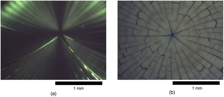
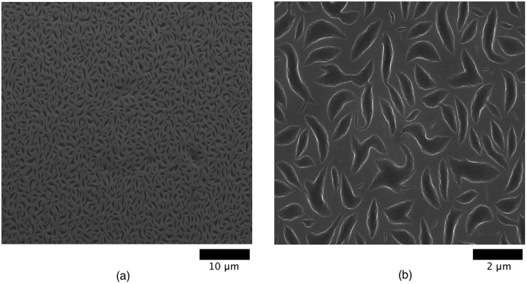
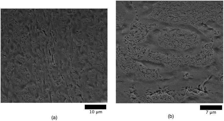
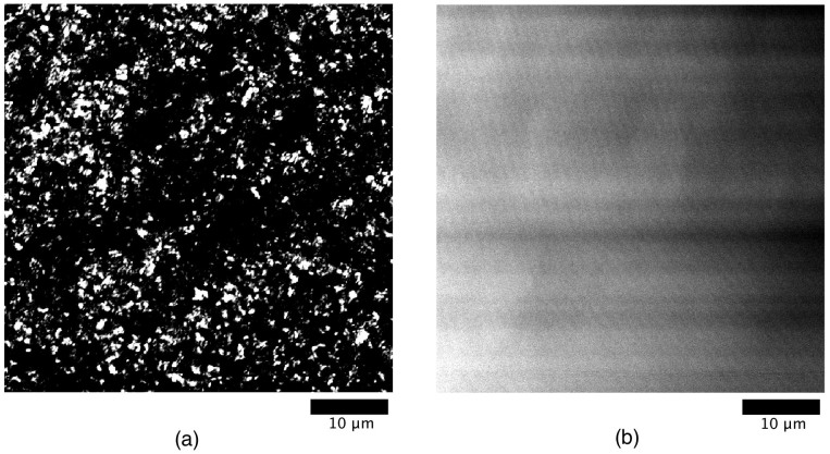
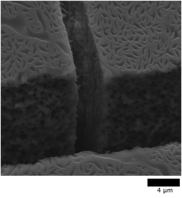
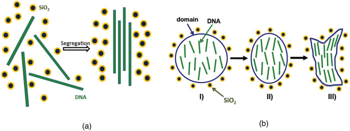
Exotic pattern formation as a result of drying of an aqueous solution containing DNA and silica nanoparticles is reported. The pattern due to segregation was found to critically depend on the relative ratio of nanoparticles and DNA, as revealed by polarization microscopy, scanning electron microscopy, and fluorescence microscopy. The blurred radial pattern that is usually observed in the drying of a colloidal solution was shown to be vividly sharpened in the presence of DNA. Uniquely curved, crescent-shaped micrometer-scale domains are generated in regions that are rich in nanoparticles. The characteristic segregated patterns observed in the present study are interpreted in terms of a large aspect ratio between the persistence length (∼ 50 nm) and the diameter (∼ 2 nm) of double-stranded DNA, and the relatively small silica nanoparticles (radius: 5 nm).
Figures
Similar articles
-
A silica nanochannel and its applications in sensing and molecular transport.Anal Chem. 2009 Jul 1;81(13):5541-8. doi: 10.1021/ac9009148. Anal Chem. 2009. PMID: 19496539
-
Preparation and characterization of chemically functionalized silica-coated magnetic nanoparticles as a DNA separator.J Phys Chem B. 2009 Jan 15;113(2):536-43. doi: 10.1021/jp807081b. J Phys Chem B. 2009. PMID: 19099431
-
Direct visual detection of DNA based on the light scattering of silica nanoparticles on a human papillomavirus DNA chip.Talanta. 2009 Dec 15;80(2):967-73. doi: 10.1016/j.talanta.2009.08.024. Epub 2009 Aug 26. Talanta. 2009. PMID: 19836580
-
Mesoporous silica nanoparticles for enhancing the delivery efficiency of immunostimulatory DNA drugs.Dalton Trans. 2014 Apr 7;43(13):5142-50. doi: 10.1039/c3dt53433b. Dalton Trans. 2014. PMID: 24496286
-
Fluorescent hybrid silica nanoparticles and their biomedical applications.Wiley Interdiscip Rev Nanomed Nanobiotechnol. 2020 May;12(3):e1603. doi: 10.1002/wnan.1603. Epub 2019 Dec 13. Wiley Interdiscip Rev Nanomed Nanobiotechnol. 2020. PMID: 31837124 Review.
Cited by
-
Exploring drying pattern of a sessile droplet of genomic DNA in the presence of hematite nanoparticles.Sci Rep. 2018 Apr 20;8(1):6352. doi: 10.1038/s41598-018-24821-1. Sci Rep. 2018. PMID: 29679031 Free PMC article.
-
A New Label-Free Technique for Analysing Evaporation Induced Self-Assembly of Viral Nanoparticles Based on Enhanced Dark-Field Optical Imaging.Nanomaterials (Basel). 2017 Dec 22;8(1):1. doi: 10.3390/nano8010001. Nanomaterials (Basel). 2017. PMID: 29271875 Free PMC article.
-
Droplets, Evaporation and a Superhydrophobic Surface: Simple Tools for Guiding Colloidal Particles into Complex Materials.Gels. 2017 May 4;3(2):15. doi: 10.3390/gels3020015. Gels. 2017. PMID: 30920512 Free PMC article. Review.
References
-
- Deegan R. D. et al. Capillary flow as the cause of ring stains from dried liquid drops. Nature 389, 827–829 (1997).
-
- Deegan R. D. Pattern formation in drying drops. Phys. Rev. E 61, 475–485 (2000). - PubMed
-
- Hampton M. A. et al. Influence of surface orientation on the organization of nanoparticles in drying nanofluid droplets. J. Colloid Interface Sci. 377, 456–462 (2012). - PubMed
-
- Weon B. M. & Je J. H. Fingering inside the coffee ring. Phys. Rev. E 87, 013003 (2013). - PubMed
-
- Parisse F. & Allain C. Drying of colloidal suspension droplets: Experimental study and profile renormalization. Langmuir 13, 3598–3602 (1997).
MeSH terms
Substances
LinkOut - more resources
Full Text Sources
Other Literature Sources
