

Review
doi: 10.1152/ajpheart.00760.2013.
Epub 2014 Jan 10.
Mechanisms of cardiac conduction: a history of revisions
Affiliations
- PMID: 24414064
- PMCID: PMC3949060
- DOI: 10.1152/ajpheart.00760.2013
Item in Clipboard
Review
Mechanisms of cardiac conduction: a history of revisions
Am J Physiol Heart Circ Physiol.
.
Abstract
Cardiac conduction is the process by which electrical excitation spreads through the heart, triggering individual myocytes to contract in synchrony. Defects in conduction disrupt synchronous activation and are associated with life-threatening arrhythmias in many pathologies. Therefore, it is scarcely surprising that this phenomenon continues to be the subject of active scientific inquiry. Here we provide a brief review of how the conceptual understanding of conduction has evolved over the last century and highlight recent, potentially paradigm-shifting developments.
Keywords: cardiac conduction; ephaptic coupling; gap junctions; modeling; myocardium.
Figures
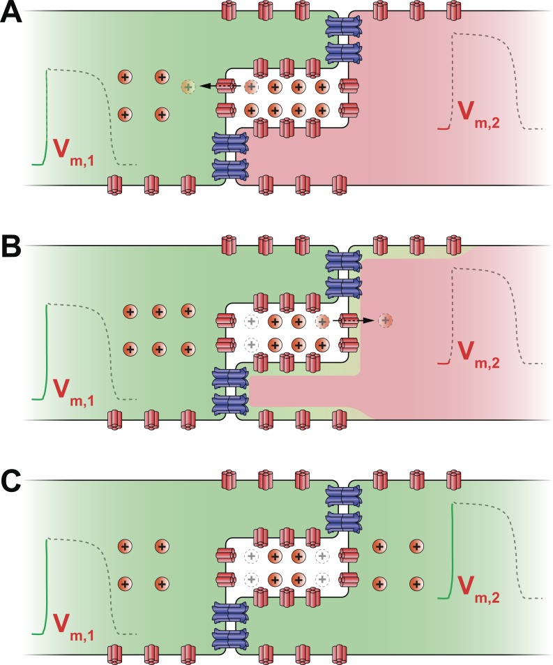
Schematic cartoon illustrating the mechanism of ephaptic coupling. A: sodium channels (shown in red) on the depolarized (green) myocyte's membrane activate and withdraw sodium ions (Na+) from the restricted extracellular cleft at the intercalated disk. As a result, the transmembrane potential (Vm,1) of the first myocyte is elevated. B: concomitant depletion of positive charge from the extracellular cleft lowers the local extracellular potential (Φe). This leads to an increase in the second, resting (red) myocyte's transmembrane potential (Vm,2), defined as the difference between its intracellular potential and the extracellular potential (Φe). In turn, sodium channels located at or near the intercalated disk of the second myocyte activate. C: sodium enters the second myocyte via these channels further depolarizing it and triggering an action potential. Thus activation is communicated ephaptically from cell to cell without the direct transfer of ions between them.
References
-
- Agullo-Pascual E, Lin X, Pfenniger A, Lubkemeier I, Willecke K, Rothenberg E, Delmar M. A novel noncanonical role of cx43 in the heart: ensuring the arrival of nav1.5 to the intercalated disk. Heart Rhythm 10: 1742, 2013
-
- Akar FG, Nass RD, Hahn S, Cingolani E, Shah M, Hesketh GG, DiSilvestre D, Tunin RS, Kass DA, Tomaselli GF. Dynamic changes in conduction velocity and gap junction properties during development of pacing-induced heart failure. Am J Physiol Heart Circ Physiol 293: H1223–H1230, 2007 - PubMed
-
- Angst BD, Khan LU, Severs NJ, Whitely K, Rothery S, Thompson RP, Magee AI, Gourdie RG. Dissociated spatial patterning of gap junctions and cell adhesion junctions during postnatal differentiation of ventricular myocardium. Circ Res 80: 88–94, 1997 - PubMed
-
- Axelsen LN, Stahlhut M, Mohammed S, Larsen BD, Nielsen MS, Holstein-Rathlou NH, Andersen S, Jensen ON, Hennan JK, Kjolbye AL. Identification of ischemia-regulated phosphorylation sites in connexin43: a possible target for the antiarrhythmic peptide analogue rotigaptide (ZP123). J Mol Cell Cardiol 40: 790–798, 2006 - PubMed
Publication types
MeSH terms
Grants and funding
LinkOut - more resources
Full Text Sources
Other Literature Sources
Miscellaneous