Comparative Study
. 2014 Mar 1;306(5):H641-53.
doi: 10.1152/ajpheart.00641.2013. Epub 2014 Jan 10.

Macrophage-derived IL-18 and increased fibrinogen deposition are age-related inflammatory signatures of vascular remodeling

Affiliations
Comparative Study

Macrophage-derived IL-18 and increased fibrinogen deposition are age-related inflammatory signatures of vascular remodeling

Luis Rodriguez-Menocal et al. Am J Physiol Heart Circ Physiol. .

Abstract

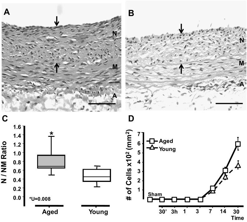
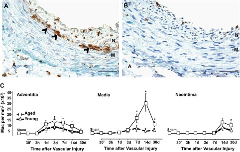
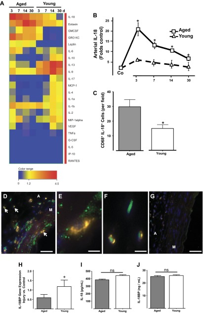
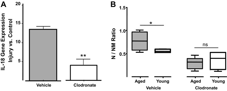
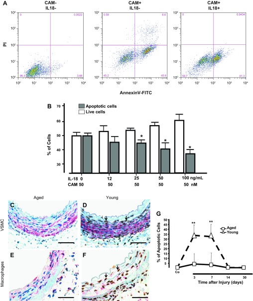
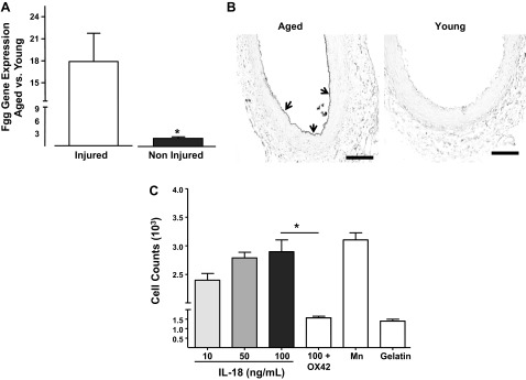
Aging has been associated with pathological vascular remodeling and increased neointimal hyperplasia. The understanding of how aging exacerbates this process is fundamental to prevent cardiovascular complications in the elderly. This study proposes a mechanism by which aging sustains leukocyte adhesion, vascular inflammation, and increased neointimal thickness after injury. The effect of aging on vascular remodeling was assessed in the rat balloon injury model using microarray analysis, immunohistochemistry, and LINCOplex assays. The injured arteries in aging rats developed thicker neointimas than those in younger animals, and this significantly correlated with a higher number of tissue macrophages and increased vascular IL-18. Indeed, IL-18 was 23-fold more abundant in the injured vasculature of aged animals compared with young rats, while circulating levels were similar in both groups of animals. The depletion of macrophages in aged rats with clodronate liposomes ameliorated vascular accumulation of IL-18 and significantly decreased neointimal formation. IL-18 was found to inhibit apoptosis of vascular smooth muscle cells (VSMC) and macrophages, thus favoring both the formation and inflammation of the neointima. In addition, injured arteries of aged rats accumulated 18-fold more fibrinogen-γ than those of young animals. Incubation of rat peritoneal macrophages with immobilized IL-18 increased leukocyte adhesion to fibrinogen and suggested a proinflammatory positive feedback loop among macrophages, VSMC, and the deposition of fibrinogen during neointimal hyperplasia. In conclusion, our data reveal that concentration changes in vascular cytokine and fibrinogen following injury in aging rats contribute to local inflammation and postinjury neointima formation.

Keywords: aging; inflammation; neointima; vascular injury; vascular smooth muscle cells.


Figures

Fig. 1.
Fig. 1.
Arteries in aged rats exhibited significantly more neointimal hyperplasia in response to injury than those of young ones. Balloon injury was inflicted in the right iliac artery of young (3–4 mo) and aged (21–24 mo) Fischer rats (n = 7/group). A and B: representative hematoxylin and eosin (H&E) sections of injured arteries from aged (A) and young (B) rats at day 30 postsurgery. Arrows mark the neointima between the internal elastic lamina and the lumen. Neointima (N), media (M), and adventitia (A) layers are noted. Scale bar = 50 μM. C: neointima formation in injured vessels from both groups at day 30 postsurgery as represented by the neointima to neointima-media thickness ratio (N/NM ratio). Statistical differences were calculated by the Mann-Whitney U-test for nonparametrically distributed data. D: temporal accumulation of cells in the neointima of injured arteries of aged and young rats. The number of cells in the noninjury, contralateral artery is labeled as control (sham). A two-tailed t-test with unequal variances (n = 4–7) was performed; *P < 0.05, significant differences.
Fig. 2.
Fig. 2.
Injured arteries accumulated more macrophages (Mac) with aging. A and B: representative microphotographs of injured arteries from aged (A) and young rats (B) harvested at 7 days postsurgery. The arrows point to CD68+-infiltrated macrophages that are stained in brown. Scale bar = 50 μM. Neointima, media, and adventitia layers are noted. C: temporal accumulation of macrophages in the neointima, media, and adventitia of injured arteries from aged and young rats. Each point in graphs represents the mean of CD68+ cell per mm2 ± SE (n = 5–10). Significant differences between cell counts in injured arteries from aged and young animals at each time point were calculated with a two-tailed t-test with unequal variances; *P < 0.05, significant values.
Fig. 3.
Fig. 3.
Aging exacerbated the accumulation of proinflammatory cytokine IL-18 at the site of vascular injury. A: heat map representing temporal cytokine variations after vascular injury in arteries from aged and young rats. Cytokines were quantified with a multiplex rat cytokine assay. Cytokine levels are expressed as fold control with respect to the cytokine concentration in the noninjured contralateral artery. GRO KC, growth-regulated oncogene-keratinocyte chemoattractant; GMCSF, granulocyte-macrophage colony-stimulating factor; MCP-1, monocyte chemotactic protein-1; G-CSF, granulocyte colony-stimulating factor; IP-10, interferon-inducible protein 10; RANTES, regulated on activation normal T-expressed and secreted. B: temporal accumulation of IL-18 in injured arteries of aged and young rats. The presurgical levels of IL-18 in both groups are labeled as control (Co). C: bar graph representing the amount of double positive macrophages for CD68 and IL-18 found in injured arteries from aged and young rats at 3 days after surgery. DG: representative microphotographs showing IL-18 (green) and CD68 (yellow) macrophages in injured arteries from aged (D) and young (G) rats harvested at 3 days after surgery. Nuclei were counterstained with DAPI (blue). Arrows point to double positive macrophages in the media and adventitia that are shown magnified in E and F, respectively. Scale bars = 50 μM (D and G) or 25 μM (E and F). Media and adventitia layers are noted. H: IL-18BP gene expression in injured arteries of aged and young rats at 3 days postsurgery determined by TaqMan qRT-PCR. IJ: plasma concentrations of IL-18 and IL-18BP in aged and young rats as determined by ELISA (n = 4/group). *P < 0.05, significant difference between groups; ns, no significant difference using a two-tailed t-test with unequal variances.
Fig. 4.
Fig. 4.
Systemic administration of clodronate liposomes decreased IL-18 accumulation in the injured vasculature while compromising neointima formation. A: gene expression of IL-18 in injured arteries of aged animals injected with either clodronate or vehicle liposomes (control) at 3 days postsurgery. Values are expressed as fold of noninjured arteries (control) ± SE. **P < 0.01, significant values; n = 4–6. B: neointima development in aged and young rats injected with either clodronate or vehicle-loaded liposomes at days −1 and +6 (n = 6–8/group). Differences between aged and young groups were calculated using a two-tailed t-test with unequal variances: *P < 0.05, significant differences; ns, nonsignificant values.
Fig. 5.
Fig. 5.
IL-18 inhibited VSMC apoptosis. A: number of apoptotic cells in rat VSMCs treated with or without IL-18 (100 ng/ml) before addition of 50 μM of camptothecin (CAM) to induce cell death. PI, propidium iodide. Early (annexin V+ PI) and late (annexin V+ PI+) apoptosis were detected by FACS analysis. Cells cultured in the absence of both IL-18 and CAM that were used as negative control for FACS analysis. B: quantitative assessment of CAM-induced apoptosis in VSMC treated with increasing concentrations of rat IL-18 (*P < 0.05; n = 4). CF: aging decreased the incidence of apoptosis during vascular remodeling. Representative microphotographs of apoptotic VSMC (C and D) and macrophages (E and F) in arteries of aged (C and E) and young (D and F) rats at 3 days after injury. Apoptotic VSMC and macrophages were detected with In Situ Oligo Ligation and immunohistochemistry for smooth muscle actin (SMA) or CD68. Apoptotic nuclei are shown in brown and cytoplasmic SMA or CD68 in red. Representative apoptotic VSMC are marked with arrowheads. Scale bars = 50 μM. G: temporal changes in the number of apoptotic cells in the vascular wall of injured arteries were represented in the plot; **P < 0.01, significant difference. The number of apoptotic cells in the noninjury, contralateral artery is labeled as control (Co).
Fig. 6.
Fig. 6.
-Immobilized IL-18 increases macrophage adhesion to fibrinogen in a Mac-1-dependent manner. A: fibrinogen γ (Fgg-γ) gene expression in aged vs. young injured arteries as measured by TaqMan RT-PCR. *P < 0.05, significant difference when n = 4. B: representative microphotography of Fgg- γ deposition in the injured artery of aged and young rats at 3 days after injury using immunohistochemistry. Fibrinogen deposition is marked with arrows. C: bar graph showing adhesion of rat peritoneal macrophages to fibrinogen-coated wells in the presence of increasing concentrations of rat IL-18. Some macrophages were allowed to adhere in the presence of OX-42, a rat Mac-1 blocking antibody. Manganese (Mn) was used as a positive control as a nonselective activator of integrin-mediated adhesion. Cells incubated in gelatin-treated wells were used as negative controls. *P < 0.05, significant differences were calculated by one-way ANOVA followed by the Bonferroni correction for multiple comparisons.

References

    1. Anderson JL, Carlquist JF. Cytokines, interleukin-18, and the genetic determinants of vascular inflammation. Circulation 112: 620–623, 2005 - PubMed
    1. Assadnia S, Rapp JP, Nestor AL, Pringle T, Cerilli GJ, Gunning WT, Webb TH, 3rd, Kligman M, Allison DC. Strain differences in neointimal hyperplasia in the rat. Circ Res 84: 1252–1257, 1999 - PubMed
    1. Chen L, Frister A, Wang S, Ludwig A, Behr H, Pippig S, Li B, Simm A, Hofmann B, Pilowski C, Koch S, Buerke M, Rose-John S, Werdan K, Loppnow H. Interaction of vascular smooth muscle cells and monocytes by soluble factors synergistically enhances IL-6 and MCP-1 production. Am J Physiol Heart Circ Physiol 296: H987–H996, 2009 - PubMed
    1. Danenberg HD, Fishbein I, Epstein H, Waltenberger J, Moerman E, Monkkonen J, Gao J, Gathi I, Reichi R, Golomb G. Systemic depletion of macrophages by liposomal bisphosphonates reduces neointimal formation following balloon-injury in the rat carotid artery. J Cardiovasc Pharmacol 42: 671–679, 2003 - PubMed
    1. Danenberg HD, Fishbein I, Gao J, Monkkonen J, Reich R, Gati I, Moerman E, Golomb G. Macrophage depletion by clodronate-containing liposomes reduces neointimal formation after balloon injury in rats and rabbits. Circulation 106: 599–605, 2002 - PubMed

Publication types

MeSH terms

LinkOut - more resources