. 2014 Mar 1;306(5):L397-404.
doi: 10.1152/ajplung.00265.2013. Epub 2014 Jan 10.

Enhanced lysis and accelerated establishment of viscoelastic properties of fibrin clots are associated with pulmonary embolism

Affiliations

Enhanced lysis and accelerated establishment of viscoelastic properties of fibrin clots are associated with pulmonary embolism

Marissa R Martinez et al. Am J Physiol Lung Cell Mol Physiol. .

Abstract

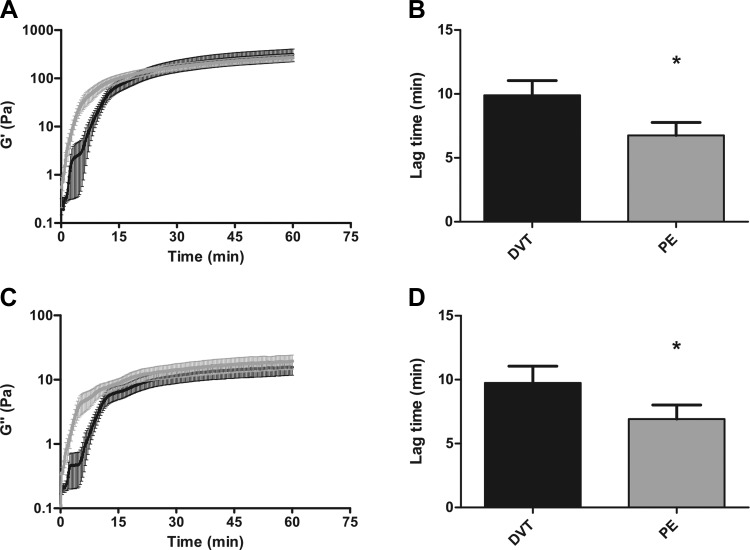
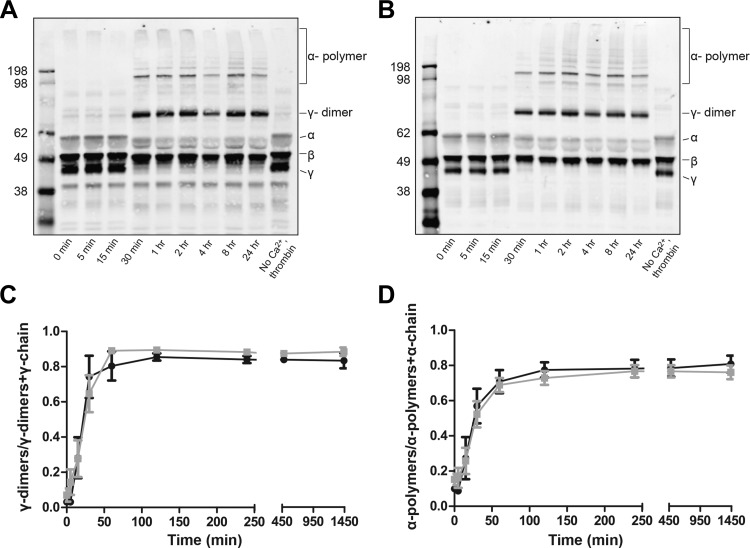
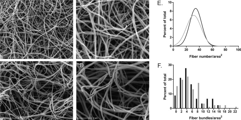
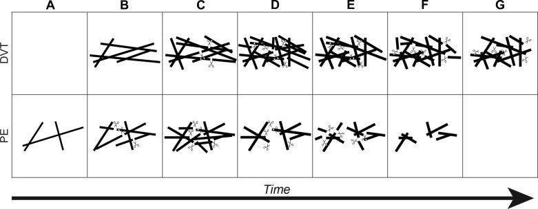
The factors that contribute to pulmonary embolism (PE), a potentially fatal complication of deep vein thrombosis (DVT), remain poorly understood. Whereas fibrin clot structure and functional properties have been implicated in the pathology of venous thromboembolism and the risk for cardiovascular complications, their significance in PE remains uncertain. Therefore, we systematically compared and quantified clot formation and lysis time, plasminogen levels, viscoelastic properties, activated factor XIII cross-linking, and fibrin clot structure in isolated DVT and PE subjects. Clots made from plasma of PE subjects showed faster clot lysis times with no differences in lag time, rate of clot formation, or maximum absorbance of turbidity compared with DVT. Differences in lysis times were not due to alterations in plasminogen levels. Compared with DVT, clots derived from PE subjects showed accelerated establishment of viscoelastic properties, documented by a decrease in lag time and an increase in the rate of viscoelastic property formation. The rate and extent of fibrin cross-linking by activated factor XIII were similar between clots from DVT and PE subjects. Electron microscopy revealed that plasma fibrin clots from PE subjects exhibited lower fiber density compared with those from DVT subjects. These data suggest that clot structure and functional properties differ between DVT and PE subjects and provide insights into mechanisms that may regulate embolization.

Keywords: deep vein thrombosis; fibrin; lysis; pulmonary embolism.


Figures

Fig. 1.
Fig. 1.
Viscoelastic properties in deep vein thrombosis (DVT) and pulmonary embolism (PE) subjects. A: formation of the clot elastic property (G′) as a function of time in DVT (black, n = 21) and PE (gray, n = 28) subjects. B: lag time is significantly faster in PE subjects compared with DVT (*P = 0.0186). C: formation of the clot viscous property (G") as a function of time in DVT and PE subjects. D: lag time is significantly faster in PE subjects compared with DVT (*P = 0.0299). Both the elastic and viscous properties are measured in Pascals (Pa). Error bars represent the SD of the mean.
Fig. 2.
Fig. 2.
Factor XIIIa cross-linking of fibrin fibers within plasma clots as a function of time in DVT and PE subjects. Representative Western blots from DVT (A) and PE (B) subjects. γ-Dimers and α-polymers both appear in DVT and PE at 30 min. The γ-chain is completely cross-linked by 1 h, whereas some α-chain remains at 24 h. C: formation of γ-dimers normalized to total γ-chain over time in DVT (black, n = 6) and PE (gray, n = 12) subjects. The rate {DVT: 0.03 [95% confidence interval (CI), 0.02–0.04] γ-dimers/min; PE: 0.02 (95% CI, 0.01–0.03) γ-dimers/min; P = 0.3332}, as determined by linear regression analysis, and extent (DVT: 0.83 ± 0. 10 γ-dimers; PE: 0.82 ± 0. 15 γ-dimers; P = 0.8809) of γ-dimer formation was similar between clots from DVT and PE subjects. D: formation of α-polymers normalized total α-chain over time in DVT and PE subjects. The rate [DVT: 0.02 (95% CI, 0.01–0.03) α-polymers/min; PE: 0.01 (95% CI, 0.01–0.02) α-polymers/min; P = 0.4216] and extent (DVT: 0.78 ± 0. 10 α-polymers; PE: 0.67 ± 0. 12 α-polymers; P = 0.0767) of α-polymer formation were similar between clots from DVT and PE subjects.
Fig. 3.
Fig. 3.
Scanning electron micrographs of clots formed from the plasma of DVT and PE subjects. A and B: representative micrographs from an individual DVT subject. Magnification bars represent 5 (A) and 2 (B) μm. C and D: representative micrographs from an individual PE subject. Magnification bars represent 5 (C) and 2 (D) μm. Bundles are indicated by arrowheads. E: nonlinear regression fit of Gaussian distributions of fiber density for DVT (black, n = 7) and PE (gray, n = 8) subjects. F: histograms for the distributions of fiber bundles in DVT (black, n = 7) and PE (gray, n = 8) subjects. Histograms for fiber density and fiber bundling were normalized to the total number of fibers or bundles, respectively, and are represented as the percent of total.
Fig. 4.
Fig. 4.
Schematic representation of clot formation and lysis in DVT and PE subjects. Fibrin fibers are denoted by solid lines and plasmin by scissors. A: in DVT clots, fibrin fibers have not formed. In PE clots, thin fibers and an initial network begin to develop. B: DVT clots now form fibrin fibers and a network. In PE clots, tissue plasminogen activator (tPA) and plasminogen begin to bind and lyse fibers. Concomitantly, new fibers are added to the network. C: new fibers are added to DVT clots, and tPA and plasminogen begin to bind and lyse fibers. New fibers continue to be added to the PE clot, and existing fibers thicken. More plasmin is generated due to additional fibrin formation. D: in DVT clots, new fibers are added to the network, existing fibers are thickened, and more plasmin is generated. The fibrin network is fully formed in both DVT and PE clots. Here, plasmin continues to lyse the clots. Fibers reach their final diameter and are comparable between DVT and PE, resulting in similar maximum turbidity during clot formation and lysis time assays. E: for both DVT and PE clots, plasmin lysis continues. However, lysis proceeds at a faster rate in PE clots due to decreased fiber density. F: lysis continues in DVT clots, whereas lysis is mostly complete in PE clots, with small fibrin fragments and fibrin degradation products produced. G: lysis advances in DVT clots, but significant clot structure remains due to increased fiber density. Plasmin has completely degraded the fibrin clot in PE subjects, and all degradation products and fibrin structures have been removed by flowing blood.

References

    1. Anderson FA, Jr, Wheeler HB, Goldberg RJ, Hosmer DW, Patwardhan NA, Jovanovic B, Forcier A, Dalen JE. A population-based perspective of the hospital incidence and case-fatality rates of deep vein thrombosis and pulmonary embolism. The Worcester DVT Study. Arch Intern Med 151: 933–938, 1991 - PubMed
    1. Bannish BE, Keener JP, Fogelson AL. Modelling fibrinolysis: a 3D stochastic multiscale model. Math Med Biol In press - PMC - PubMed
    1. Carr ME, Jr, Hermans J. Size and density of fibrin fibers from turbidity. Macromolecules 11: 46–50, 1978 - PubMed
    1. Collet JP, Allali Y, Lesty C, Tanguy ML, Silvain J, Ankri A, Blanchet B, Dumaine R, Gianetti J, Payot L, Weisel JW, Montalescot G. Altered fibrin architecture is associated with hypofibrinolysis and premature coronary atherothrombosis. Arterioscler Thromb Vasc Biol 26: 2567–2573, 2006 - PubMed
    1. Collet JP, Park D, Lesty C, Soria J, Soria C, Montalescot G, Weisel JW. Influence of fibrin network conformation and fibrin fiber diameter on fibrinolysis speed: dynamic and structural approaches by confocal microscopy. Arterioscler Thromb Vasc Biol 20: 1354–1361, 2000 - PubMed

Publication types

MeSH terms

LinkOut - more resources