. 2014 Apr;16(4):493-504.
doi: 10.1093/neuonc/not242. Epub 2014 Jan 10.

mTORC1 inhibition delays growth of neurofibromatosis type 2 schwannoma

Affiliations

mTORC1 inhibition delays growth of neurofibromatosis type 2 schwannoma

Marco Giovannini et al. Neuro Oncol. 2014 Apr.

Abstract

Background: Neurofibromatosis type 2 (NF2) is a rare autosomal dominant genetic disorder, resulting in a variety of neural tumors, with bilateral vestibular schwannomas as the most frequent manifestation. Recently, merlin, the NF2 tumor suppressor, has been identified as a novel negative regulator of mammalian target of rapamycin complex 1 (mTORC1); functional loss of merlin was shown to result in elevated mTORC1 signaling in NF2-related tumors. Thus, mTORC1 pathway inhibition may be a useful targeted therapeutic approach.

Methods: We studied in vitro cell models, cohorts of mice allografted with Nf2(-/-) Schwann cells, and a genetically modified mouse model of NF2 schwannoma in order to evaluate the efficacy of the proposed targeted therapy for NF2.

Results: We found that treatment with the mTORC1 inhibitor rapamycin reduced the severity of NF2-related Schwann cell tumorigenesis without significant toxicity. Consistent with these results, in an NF2 patient with growing vestibular schwannomas, the rapalog sirolimus induced tumor growth arrest.

Conclusions: Taken together, these results constitute definitive evidence that justifies proceeding with clinical trials using mTORC1-targeted agents in selected patients with NF2 and in patients with NF2-related sporadic tumors.

Keywords: neurofibromatosis type 2; rapamycin; schwannoma.


Figures
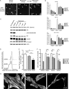
Fig. 1.
Fig. 1.
Rapamycin inhibits growth and reduces cell size of human and murine NF2−/− schwannoma cells. (A and B) Soft agar colony formation assay was performed with the HEI193 human schwannoma cell line, the 08031-9 mouse schwannoma cell line, and ESC-FC1801 mouse Nf2−/− Schwann cells. After 3 weeks of treatment, the number of colonies was evaluated for the different concentrations of rapamycin or vehicle treatment, and reduction in colony formation was shown with rapamycin treatment. Then, the same plates were incubated without rapamycin for an additional 3 weeks: the increased number of colonies demonstrated a cytostatic effect of rapamycin. (C) The 08031-9 mouse schwannoma cell line was treated with different concentrations of rapamycin or with vehicle or not treated for 24 h. Western blot analysis showed strong inhibition of S6 and 4E-BP1 phosphorylation without increase of Akt phosphorylation. p-Akt, phosphorylated Akt. (D and E) Mouse Nf2−/− Schwann cells were treated with rapamycin at 20 nM and 20 µM, harvested, stained with propidium iodide, and analyzed by flow cytometry to determine FSC-H of G1-phase cells. (D) Shift of FSC-H with rapamycin treatment for one experiment. (E) FSC-H average for 3 independent experiments. (F and G) Primary cultures of human schwannoma cells were treated with rapamycin at 20 nM, 40 nM, or 400 nM for 12–20 days then fixed and stained for S100 protein (G). The cell surface area of S100 protein–positive cells was assessed with an automated Celigo Cell Cytometer. The 4 VS cell cultures showed a reduction of the mean cell area after rapamycin treatment (F). Data on VS234 were not the result of performance in triplicate, therefore statistical significance is not provided. Error bar of VS234 cells treated with 20 nM are relative to duplicate wells.
Fig. 2.
Fig. 2.
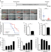
Rapamycin rapidly inhibited tumor growth and improved functional outcomes in an orthotopic NF2 mouse schwannoma allograft model. (A) Nu/nu mice were implanted with adult mouse Nf2−/− Schwann cells 1 week before randomization. Mice were treated daily with rapamycin (8 mg/kg/d, i.p.) (n = 20) or vehicle (n = 15) for 14 days. (B and C) Rapamycin treatment reduced the growth of intraneural Nf2−/− Schwann cell allografts by 4-fold compared with vehicle-treated controls (P = .001 at day 22) and (D) significantly delayed the onset of severe hind-limb paralysis. (E and F) Rapamycin treatment reduced the number of BrdU-positive cells (E) and microvascular density (F) in tumors. (G–I) Quantification of absorbance for phospho-S6 protein normalized to total S6 by ELISA demonstrated decreased phosphorylation at 1 h, 6 h, and 16 h following i.p. administration of 8 mg/kg/d rapamycin (G). Maximum inhibition was observed at the 1-h time point. Phospho–4E-BP1 levels normalized to α-tubulin were also decreased, confirming inhibition of the mTOR pathway 1 h after drug administration (H). However, likely due to the existence of a negative feedback loop emanating from S6K,, we noted that rapamycin transiently activated Akt (I).
Fig. 3.
Fig. 3.
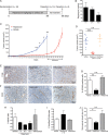
Rapamycin suppresses the growth of a transplantable mouse schwannoma model. (A) Mice bearing 08031-9 tumors were treated with rapamycin 8 mg/kg/d for 5/7 days per week for 36 days. For 3 mice, treatment was discontinued for 14 days before sacrifice. (B) Pharmacokinetic analysis of rapamycin-treated mice showed that rapamycin concentration levels in tumors reached levels consistent with activity in vitro. (C) Rapamycin potently suppressed schwannoma growth, showing potent cytostatic effects (P = .0004 at the endpoint, Mann–Whitney test). Three mice in the treated group were kept without treatment to evaluate the effect of rapamycin withdrawal on tumor growth, showing that rapamycin-induced inhibition of tumor cell proliferation was reversible. (D) After 36 days of treatment, the rapamycin-treated group demonstrated reduction in tumor volume >5-fold compared with controls (P < .001, t-test). (E–J) Rapamycin treatment inhibited mTOR activity and activated Akt. (E) Phospho-S6 protein immunohistochemistry in vehicle- and rapamycin-treated tumors showed decreased phosphorylation at 1 h after rapamycin administration. (F) CD31 immunohistochemistry highlights abnormal vascular morphology and increased vessel diameter in the vehicle and withdrawal groups. Tumor vessel morphology was improved in the rapamycin-treated group. (G) Quantification of BrdU incorporation demonstrated significant reduction of proliferating tumor cells following rapamycin treatment (P = .0018 vs vehicle). Fourteen days after treatment withdrawal, the proliferating index regained the control level. (H) Quantification of absorbance for phospho-S6 protein normalized to total S6 by ELISA showed decreased phosphorylation at 1 h, 6 h, and 16 h. Maximum inhibition was observed at the 6-h time point. (I) As a result of mTOR inhibition, densitometry-quantified levels of phospho-Akt normalized to total Akt showed activation 1 h after the last rapamycin dose. (J) Quantification of intratumoral vascular density (iMVD) evaluated in vehicle (n = 6), rapamycin (n = 4), and withdrawal (n = 3) groups demonstrated a significantly decreased iMVD in the treated group (P = .012 vs vehicle). Increased iMVD was observed after drug withdrawal (P = ns vs vehicle).
Fig. 4.
Fig. 4.
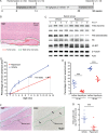
Rapamycin inhibits tumor growth in an NF2 schwannoma genetically engineered mouse model. (A) Starting at 6 weeks of age, transgenic P0-SCH-Δ(39-121)-27 mice were randomized and treated daily with rapamycin (8 mg/kg/d, i.p., 5/7 d/wk) (n = 14) or vehicle (n = 15) for 8 weeks. After 8 weeks of treatment, 8 mice/group were analyzed, while the remaining mice (n = 13) were treated following a weekly regimen of rapamycin (16 mg/kg/d, i.p., 1/7 d/wk) or vehicle until 50 weeks old, when the initial drug regimen was resumed for 6 additional weeks. (B) For each mouse, the surface ratio between tumoral and normal nerve tissue was calculated. H&E, hematoxylin and eosin. (C) Transgenic P0-SCH-Δ(39-121)-27 mice were treated during 7 days with rapamycin (8 mg/kg/d) or vehicle. Spinal nerves were collected 1 h after the last dose, and protein was analyzed by western blot, showing decreased phosphorylation of S6 and 4E-BP1 proteins and a slight increase in Akt phosphorylation. (D) Natural history of Schwann cell tumor development in the transgenic P0-SCH-Δ(39-121)-27 mice. The maintenance regimen delayed the growth of spinal root tumors compared with the predicted tumor burden in untreated mice. (E) Average tumor burden ± SEM in the spinal roots (40 ± 4 per mouse) is presented as a tumor ratio at the end of 14 weeks and 56 weeks of treatment (P < .001, multilevel analysis). (F) BrdU injections were performed in rapamycin- and vehicle-treated mice. The green line shows the nerve root area where the BrdU-positive cells were counted. H&E, hematoxylin and eosin. (G) Quantification of BrdU-positive cells showing reduction of proliferation in rapamycin-treated mice (56 wk treatment).
Fig. 5.
Fig. 5.
Volumetric evolution of VS in an NF2 patient treated with sirolimus. (A) Annual volumetric tumor growth of the right VS decreased under sirolimus therapy from 47% per year to 3.5% per year. Sirolimus blood dosages are indicated in the lower right part of the graph. (B) T1 axial gadolinium-enhanced sequences showing the target VS at 4 different time points during evolution (identical scale bar in each MRI = 1 cm). The table shows the volume of VS and the corresponding cystic component during sirolimus therapy.

Comment in

References

    1. Dolecek TA, Propp JM, Stroup NE, et al. CBTRUS statistical report: primary brain and central nervous system tumors diagnosed in the United States in 2005–2009. Neuro Oncol. 2012;14(Suppl 5):v1–v49. - PMC - PubMed
    1. Evans DG, Huson SM, Donnai D, et al. A clinical study of type 2 neurofibromatosis. Q J Med. 1992;84:603–618. - PubMed
    1. Blakeley JO, Evans DG, Adler J, et al. Consensus recommendations for current treatments and accelerating clinical trials for patients with neurofibromatosis type 2. Am J Med Genet A. 2012;158A:24–41. - PMC - PubMed
    1. Hadfield KD, Smith MJ, Urquhart JE, et al. Rates of loss of heterozygosity and mitotic recombination in NF2 schwannomas, sporadic vestibular schwannomas and schwannomatosis schwannomas. Oncogene. 2010;29:6216–6221. - PubMed
    1. Stemmer-Rachamimov AO, Xu L, Gonzalez-Agosti C, et al. Universal absence of merlin, but not other ERM family members, in schwannomas. Am J Pathol. 1997;151:1649–1654. - PMC - PubMed

Publication types

MeSH terms