. 2014 Jul;17(3):617-30.
doi: 10.1007/s10456-014-9414-9. Epub 2014 Jan 11.

Endothelial cells expressing low levels of CD143 (ACE) exhibit enhanced sprouting and potency in relieving tissue ischemia

Affiliations

Endothelial cells expressing low levels of CD143 (ACE) exhibit enhanced sprouting and potency in relieving tissue ischemia

Eduardo A Silva et al. Angiogenesis. 2014 Jul.

Abstract

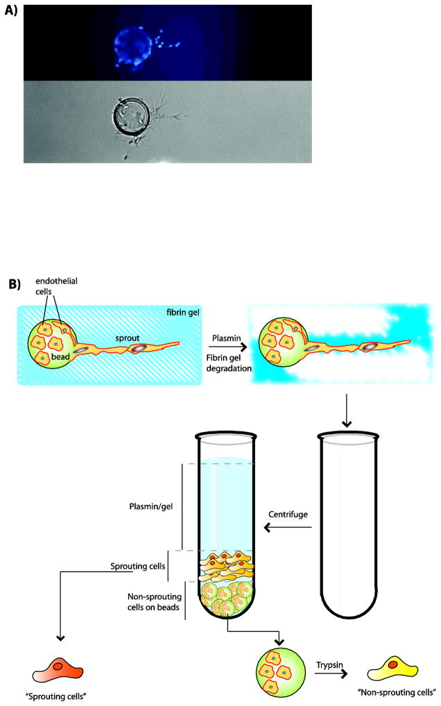
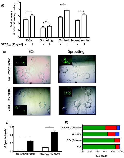
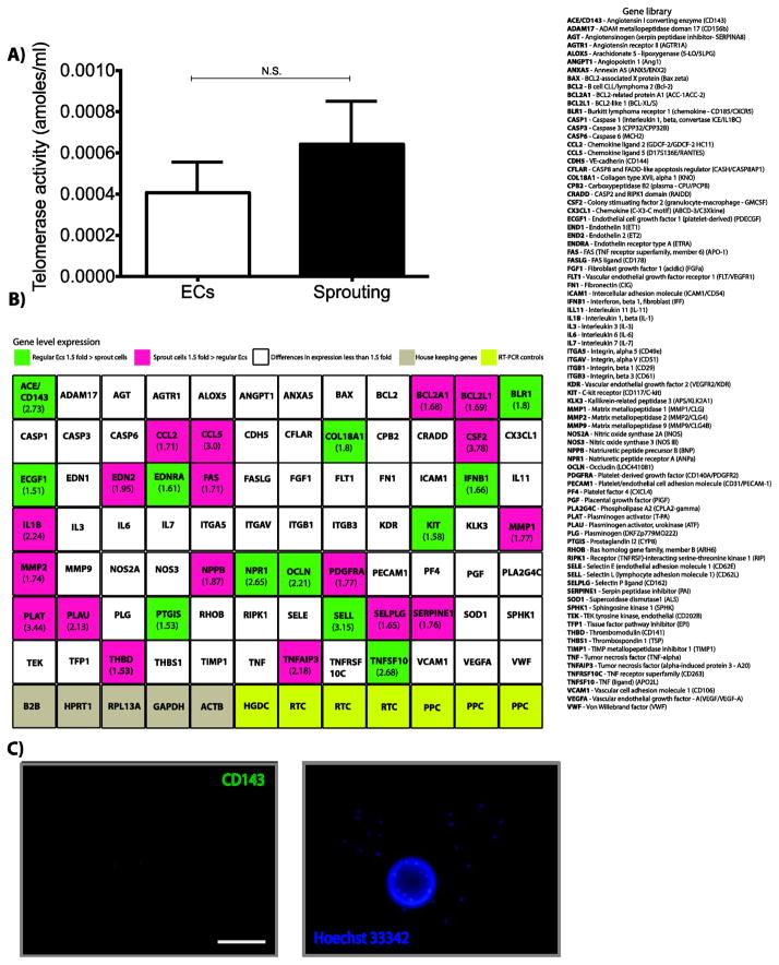
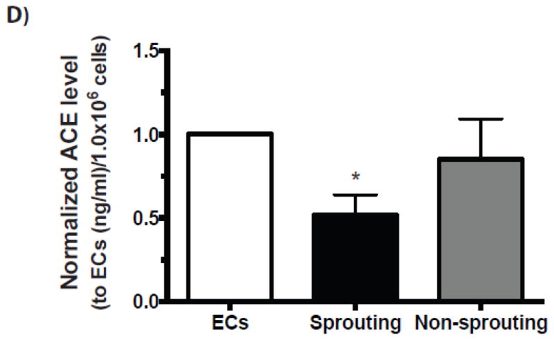
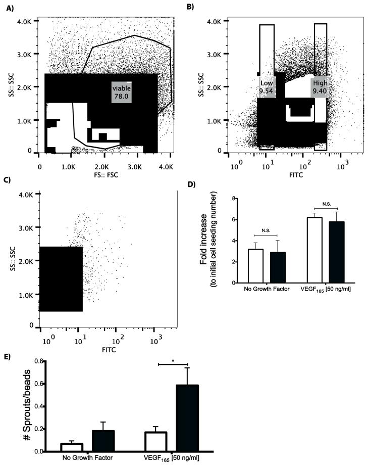
The sprouting of endothelial cells from pre-existing blood vessels represents a critical event in the angiogenesis cascade. However, only a fraction of cultured or transplanted endothelial cells form new vessels. Moreover, it is unclear whether this results from a stochastic process or instead relates to certain endothelial cells having a greater angiogenic potential. This study investigated whether there exists a sub-population of cultured endothelial cells with enhanced angiogenic potency in vitro and in vivo. First, endothelial cells that participated in sprouting, and non-sprouting cells, were separately isolated from a 3D fibrin gel sprouting assay. Interestingly, the sprouting cells, when placed back into the same assay, displayed a sevenfold increase in the number of sprouts, as compared to control cells. Angiotensin-converting enzyme (CD143) was significantly down regulated on sprouting cells, as compared to regular endothelial cells. A subset of endothelial cells with low CD143 expression was then prospectively isolated from an endothelial cell culture. Finally, these cells were found to have greater potency in alleviating local ischemia, and restoring regional blood perfusion when transplanted into ischemic hindlimbs, as compared to unsorted endothelial cells. In summary, this study indicates that low expression of CD143 can be used as a biomarker to identify an endothelial cell sub-population that is more capable to drive neovascularization.


Figures

Figure 1
Figure 1
Depiction of endothelial cell sprouts formed in a fibrin based gel sprouting assay. (A) Fluorescent (top) and phase-contrast (bottom) photomicrographs of HMVEC-D cells sprouting from microcarriers at day 3. Cells were labeled with DAPI for nucleus localization and further cell quantification. (B) Diagram describing the approach utilized to isolate HMVEC-d cells that formed sprouts (“sprouting cell”) from cells that remained on the microcarriers (“non sprouting cell”). A combination of fibrin gel degradation mediated by plasmin, and centrifugation was utilized to separate the sprouting cells from the cells remaining adherent to the microcarrier beads. Non-sprouting cells were subsequently removed from microcarrier beads using trypsinization. Photomicrographs in 1A are at 200x.
Figure 2
Figure 2
Characterization of sprouting and non-sprouting cells. (A) Proliferation of cells isolated from sprouts (Sprouting) and cells which remained on the beads (Non-sprouting), as compared with regular HMVEC-d (ECs) (not placed in fibrin gels) and HMVEC-d cells that were cultured in the fibrin gels and were subject to the exact same process of isolation as the sprouting and non-sprouting cells (Control). All cell types were tested in media containing either no growth factors, or this media supplemented with VEGF165 [50 ng/mL]. An equal number of each type of cell was seeded in each well, and the resultant cell number at three days quantified. (B) Representative fluorescent (inserts) and phase-contrast photomicrographs displaying the density and quantity of sprouts formed both from the regular HMVEC-d (ECs) and the sprouting cells under both cell media containing VEGF and no growth factors. (C) Cells that formed sprouts (sprouting cells; dark bars) displayed a considerable greater ability to make new sprouts when placed back into the assay, as compared with regular HMVEC-d (white filled bars), both under VEGF165 supplemented media and no growth factor media. (D) Histogram of the distribution of number of sprouts formed by regular ECs (both observed and theoretical – Poisson values) and Sprouting cells (both observed and theoretical – Poisson values) for microcarriers displaying zero sprouts (green stack bar), one sprout (red stack bar), two sprouts (blue stack bar), three sprouts (grey stack bar), four sprouts (orange stack bar). Mean values are presented with standard deviations, * indicates statistically significant differences (p<0.05) between conditions and N.S. displays no statistically significant difference between conditions. All photomicrographs (insert included) in 2B are at 200x, and calibration bar represents 100 μm.
Figure 3
Figure 3
Characterization of sprouting cells. (A) Telomerase activity of control HMVEC-d endothelial cells (ECs) as compared with HMVEC-d endothelial cells that had previously formed sprouts (Sprouting). (B) Expression levels of eighty-four signature angiogenic genes in control HMVEC-d and HMVEC-d that formed sprouts. The number in parenthesis represents the fold change between the two cell populations, and each gene is color coded to represent the magnitude of change and whether an increase or decrease was noted with sprouting cells. (C) Microcarriers seeded with HMVEC-d were embedded in a 3D fibrin gel and fed daily for 5 days with EGM-2MV. After fixation, gels were incubated with a nuclear stain (Hoechst 33342 – right panel) and CD143 antibody (left panel). Differential CD143 staining was observed between sprouting cells and cells residing on the microcarriers (non-sprouting cells). (D) The levels of ACE/CD143 in conditioned medium from HMVEC-d endothelial cells (ECs), sprouting cells and non-sprouting cells. Mean values are presented with standard deviations. * indicates statistically significant differences (p<0.05) between ECs and non-sprouting cells (E) and N.S. displays no statistically significant difference between conditions (A). Calibration bar represents 200 μm.
Figure 3
Figure 3
Characterization of sprouting cells. (A) Telomerase activity of control HMVEC-d endothelial cells (ECs) as compared with HMVEC-d endothelial cells that had previously formed sprouts (Sprouting). (B) Expression levels of eighty-four signature angiogenic genes in control HMVEC-d and HMVEC-d that formed sprouts. The number in parenthesis represents the fold change between the two cell populations, and each gene is color coded to represent the magnitude of change and whether an increase or decrease was noted with sprouting cells. (C) Microcarriers seeded with HMVEC-d were embedded in a 3D fibrin gel and fed daily for 5 days with EGM-2MV. After fixation, gels were incubated with a nuclear stain (Hoechst 33342 – right panel) and CD143 antibody (left panel). Differential CD143 staining was observed between sprouting cells and cells residing on the microcarriers (non-sprouting cells). (D) The levels of ACE/CD143 in conditioned medium from HMVEC-d endothelial cells (ECs), sprouting cells and non-sprouting cells. Mean values are presented with standard deviations. * indicates statistically significant differences (p<0.05) between ECs and non-sprouting cells (E) and N.S. displays no statistically significant difference between conditions (A). Calibration bar represents 200 μm.
Figure 4
Figure 4
FACS analysis and sorting of CD143 high and low expressing HMVEC-d, and the proliferation and sprouting of CD143 low cells. (A) Cells were stained with CD143 antibodies, and first subjected to initial gating for the viable cell population using forward scatter (FS) and side scatter (SS); Black outline on plot represents gating for viable cell population. (B) Viable cells were then sorted for the 10% of cells with the highest and lowest expression of CD143. Black rectangles represent these two cell populations. (C) HMVEC-d were stained with isotype control antibodies matching the host species of the primary antibody allowing an assessment of the level of background staining. (D) Proliferation of endothelial cells sorted for CD143 low (dark bars) as compared with regular (non-sorted) endothelial cells (white bars) under media containing either no growth factors or media supplemented with VEGF165 [50 ng/mL]. (E) Sprouting of endothelial cells expressing low levels of CD143 (black bars), as compared to regular HMVEC-d (white filled bars), in media with no growth factors or media that contains no growth factors except VEGF165. Mean values are presented with standard deviations. N.S. indicates no statistically significant difference between conditions and * indicates statistically significant differences (p<0.05) between conditions.
Figure 5
Figure 5
The ability of CD143 low expression endothelial cells to drive neovascularization in murine ischemic hindlimbs was compared with regular (unsorted) endothelial cells. (A) Representative images of muscle tissue stained for the endothelial cell marker CD31 following transplantation of regular endothelial cells, or CD143 low cells at 6 weeks post-treatment. (B) The blood vessel density in muscle tissue was quantified for these two experimental conditions. (C) Perfusion of ischemic hindlimbs over time following transplantation of CD143 low cells (●) or regular endothelial cells (□). (D) The distribution of severity of ischemia was monitored over time for both the animals treated with CD143 low cells and the animals treated with regular endothelial cells. Ischemia was graded as no necrosis (□), toe discoloration ( formula image
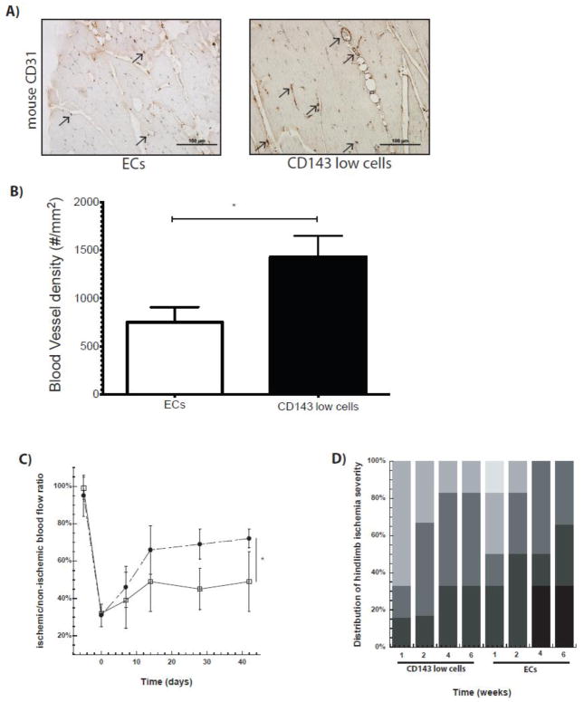
), one necrotic toe ( formula image), two or more necrotic toes ( formula image), necrotic foot ( formula image) and autoamputation (■). Mean values are presented with standard deviations, * indicates statistically significant differences (p<0.05) between conditions. Arrows in (A) highlight positive CD31 cells.

Similar articles

Cited by

References

    1. Carmeliet P, Jain R. Molecular mechanisms and clinical applications of angiogenesis. Nature. 2011;473 (7347):298–307. doi: 10.1038/nature10144. - DOI - PMC - PubMed
    1. Semenza G. Oxygen sensing, homeostasis, and disease. The New England journal of medicine. 2011;365 (6):537–547. doi: 10.1056/NEJMra1011165. - DOI - PubMed
    1. Potente M, Gerhardt H, Carmeliet P. Basic and therapeutic aspects of angiogenesis. Cell. 2011;146 (6):873–887. doi: 10.1016/j.cell.2011.08.039. - DOI - PubMed
    1. Davis G, Senger D. Endothelial extracellular matrix: biosynthesis, remodeling, and functions during vascular morphogenesis and neovessel stabilization. Circ Res. 2005;97 (11):1093–1107. doi: 10.1161/01.RES.0000191547.64391.e3. - DOI - PubMed
    1. Ribatti D, Crivellato E. “Sprouting angiogenesis”, a reappraisal. Developmental biology. 2012;372 (2):157–165. doi: 10.1016/j.ydbio.2012.09.018. - DOI - PubMed

Publication types

LinkOut - more resources