Review
. 2014 Mar;196(6):1133-42.
doi: 10.1128/JB.01155-13. Epub 2014 Jan 10.

Lipoteichoic acids, phosphate-containing polymers in the envelope of gram-positive bacteria

Affiliations
Review

Lipoteichoic acids, phosphate-containing polymers in the envelope of gram-positive bacteria

Olaf Schneewind et al. J Bacteriol. 2014 Mar.

Abstract

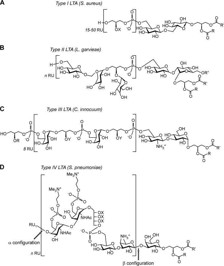
Lipoteichoic acids (LTA) are polymers of alternating units of a polyhydroxy alkane, including glycerol and ribitol, and phosphoric acid, joined to form phosphodiester units that are found in the envelope of Gram-positive bacteria. Here we review four different types of LTA that can be distinguished on the basis of their chemical structure and describe recent advances in the biosynthesis pathway for type I LTA, d-alanylated polyglycerol-phosphate linked to di-glucosyl-diacylglycerol. The physiological functions of type I LTA are discussed in the context of inhibitors that block their synthesis and of mutants with discrete synthesis defects. Research on LTA structure and function represents a large frontier that has been investigated in only few Gram-positive bacteria.


Figures

FIG 1
FIG 1
Structure of LTA molecules from various bacteria. The repeating unit, RU, of each LTA type is indicated within the brackets; it is always linked to a glycolipid. Where known, n (the number of RUs) is indicated. (A) Structure of type I LTA from Staphylococcus aureus. The 1,3-polyglycerol-phosphate RUs are substituted at the C2 position (X) with hydrogen proton (∼15%), d-alanyl ester (∼70%), or N-acetylglucosamine (∼15%). (B) Structure of type II LTA from Lactococcus garvieae. (C) Structure of type III LTA from Clostridium innocuum. Gro-P in the RU can be substituted at the C2 position (Y) with hydrogen proton (∼25%), glucosamine (∼50%), or N-acetylglucosamine (∼50%). (D) Structure of type IV LTA of Streptococcus pneumoniae. The RU may be substituted with hydrogen, d-alanyl, or N-acetylglucosamine (X). Substituents R, R′, and R″ in the glycolipids may be alkyl or branched alkyl chains.
FIG 2
FIG 2
UDP-glucose contributes to wall teichoic acid and lipoteichoic acid synthesis. The diagram shows the flow of the UDP-glucose (UDP-Glc) pool in Bacillus subtilis and the key enzymes involved in synthesis of lipoteichoic acid (LTA) as well as minor and major wall teichoic acids (WTA) (adapted from reference 62). The major WTA from B. subtilis 168 is d-alanyl-[α-d-glycosylated poly(Gro-P)]. It is replaced with a phosphorus-free polysaccharide containing uronic acid residues (minor WTA) when B. subtilis is grown in phosphate-limited medium (89, 137). See the text for details.
FIG 3
FIG 3
Synthesis of lipoteichoic acid in Staphylococcus aureus. (A) Diagram showing the reaction catalyzed by LtaS. The first glycerophosphate (Gro-P) subunit is cleaved from phosphatidyl glycerol (PG) and attached to the glycolipid anchor diglycosyl-diacylglycerol (Glc2DAG). This reaction leads to the release of DAG. Polymerization of Gro-P by LtaS occurs at the distal end of Glc2DAG-(Gro-P)1, utilizing additional PG molecules (n = ∼50). For some bacterial species, the first reaction is thought to require a specific primase (see the text for details). The DgkB enzyme is responsible for the recycling of DAG (see the text for details). (B) Diagram showing the biosynthesis of LTA in S. aureus. The enzymes PgcA, GtaB, and YpfP synthesize the Glc2DAG glycolipid anchor. LtaA, a 12-transmembrane domain protein (in gray), flips Glc2DAG across the plasma membrane. LtaS (red) spans the plasma membrane five times, and its C-terminal domain polymerizes the poly(Gro-P) chain on Glc2DAG on the trans side of the membrane.
FIG 4
FIG 4
Models for d-alanylation of teichoic acids. DltA (Dcl), DltB, DltC (Dcp), and DltD are required for the d-alanylation of LTA, and two models have been proposed to explain the molecular basis. Model 1 proposes that the combined activity of DltA and DltC in the cytoplasm results in the transfer of d-Ala onto the lipid carrier bactoprenol pyrophosphate (PP) (95). Next, DltB flips d-Ala-bactoprenol-PP across the plasma membrane. It is unclear whether DltD assists the DltB flippase or contributes to d-Ala transfer onto LTA (96). In model 2, DltD facing the cytoplasm assists DltA for the loading of d-Ala onto the carrier protein DltC. Next, DltC-S-d-Ala is translocated across the membrane by DltB and transfers d-Ala onto LTA (89). d-Alanylation of WTA is not thought to require catalysis. Presumably, d-Ala moieties of LTA are transferred onto WTA. MurNAc, N-acetylmuramic acid; GlcNAc, N-acetylglucosamine; ManNAc, N-acetylmannosamine.

References

    1. Baddiley J, Buchanan JG, Carss B, Mathias AP. 1956. Cytidine diphosphate ribitol. Biochim. Biophys. Acta 21:191–192. 10.1016/0006-3002(56)90123-8 - DOI - PubMed
    1. Baddiley J, Buchanan JG, Carss B, Mathias AP, Sanderson AR. 1956. The isolation of cytidine diphosphate glycerol, cytidine diphosphate ribitol and mannitol 1-phosphate from Lactobacillus arabinosus. Biochem. J. 64:599–603 - PMC - PubMed
    1. Leloir LF. 1951. The enzymatic transformation of uridine diphosphate glucose into a galactose derivative. Arch. Biochem. Biophys. 33:186–190. 10.1016/0003-9861(51)90096-3 - DOI - PubMed
    1. Glaser L. 1957. The enzymic synthesis of cellulose by Acetobacter xylinum. Biochim. Biophys. Acta 25:436. 10.1016/0006-3002(57)90501-2 - DOI - PubMed
    1. Leloir LF, Cardini CE. 1957. Biosynthesis of glycogen from uridine diphosphate glucose. J. Am. Chem. Soc. 79:6340–6341. 10.1021/ja01580a061 - DOI

Publication types

MeSH terms

LinkOut - more resources