. 2014 Feb 28;289(9):6110-9.
doi: 10.1074/jbc.M113.524801. Epub 2014 Jan 12.

Physical interaction between bacterial heat shock protein (Hsp) 90 and Hsp70 chaperones mediates their cooperative action to refold denatured proteins

Affiliations

Physical interaction between bacterial heat shock protein (Hsp) 90 and Hsp70 chaperones mediates their cooperative action to refold denatured proteins

Hitoshi Nakamoto et al. J Biol Chem. .

Abstract

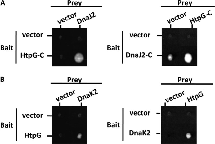
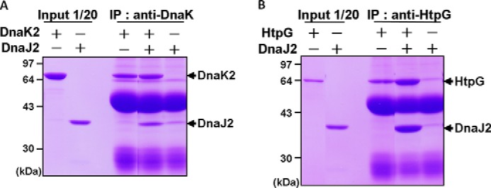
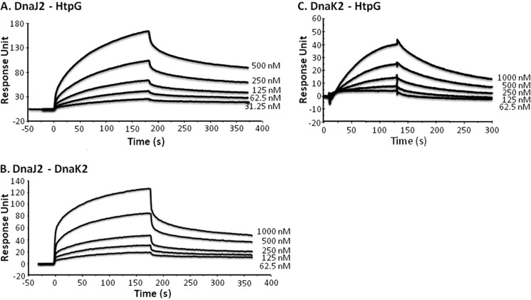
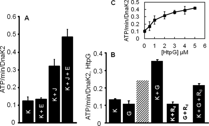
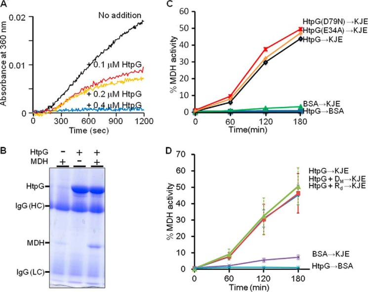
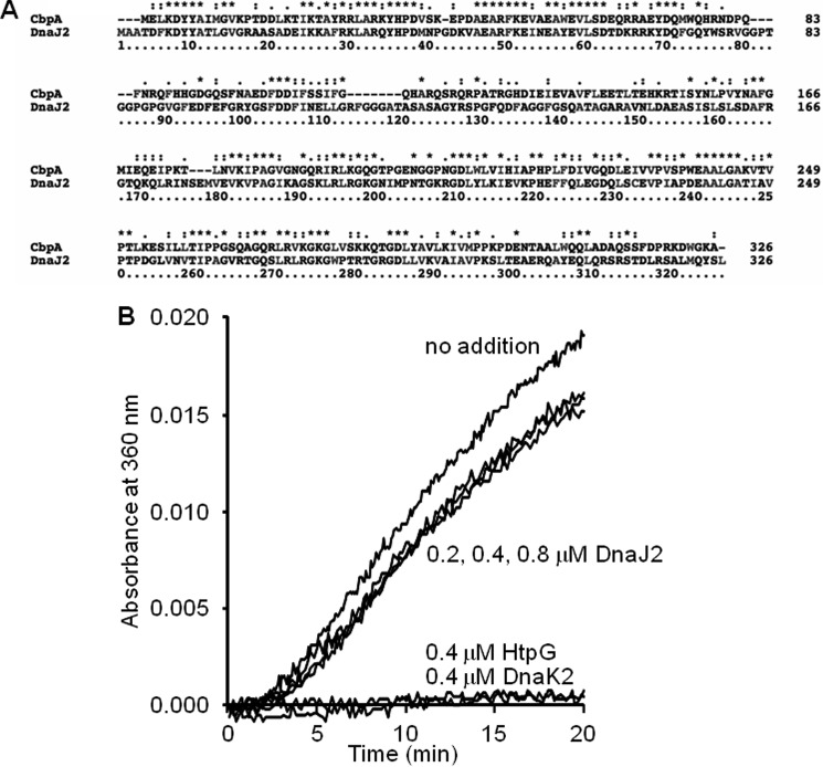
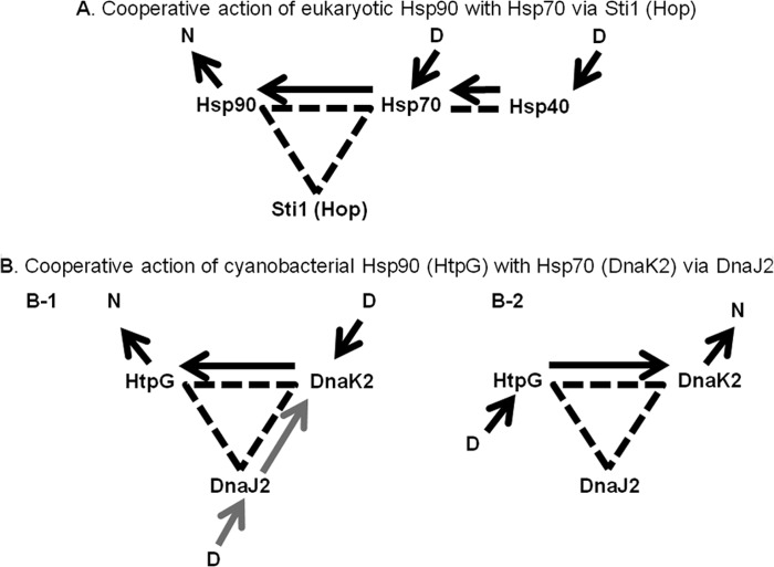
In eukaryotes, heat shock protein 90 (Hsp90) is an essential ATP-dependent molecular chaperone that associates with numerous client proteins. HtpG, a prokaryotic homolog of Hsp90, is essential for thermotolerance in cyanobacteria, and in vitro it suppresses the aggregation of denatured proteins efficiently. Understanding how the non-native client proteins bound to HtpG refold is of central importance to comprehend the essential role of HtpG under stress. Here, we demonstrate by yeast two-hybrid method, immunoprecipitation assays, and surface plasmon resonance techniques that HtpG physically interacts with DnaJ2 and DnaK2. DnaJ2, which belongs to the type II J-protein family, bound DnaK2 or HtpG with submicromolar affinity, and HtpG bound DnaK2 with micromolar affinity. Not only DnaJ2 but also HtpG enhanced the ATP hydrolysis by DnaK2. Although assisted by the DnaK2 chaperone system, HtpG enhanced native refolding of urea-denatured lactate dehydrogenase and heat-denatured glucose-6-phosphate dehydrogenase. HtpG did not substitute for DnaJ2 or GrpE in the DnaK2-assisted refolding of the denatured substrates. The heat-denatured malate dehydrogenase that did not refold by the assistance of the DnaK2 chaperone system alone was trapped by HtpG first and then transferred to DnaK2 where it refolded. Dissociation of substrates from HtpG was either ATP-dependent or -independent depending on the substrate, indicating the presence of two mechanisms of cooperative action between the HtpG and the DnaK2 chaperone system.

Keywords: Cyanobacteria; DnaJ; DnaK; Heat Shock Protein; Hsp70; Hsp90; HtpG; Molecular Chaperone; Protein Aggregation.


Figures

FIGURE 1.
FIGURE 1.
Two-hybrid interactions of HtpG with DnaJ2 and DnaK2. Yeast cells were grown for 1 week at 30 °C on an SC plate lacking leucine, tryptophan, and adenine. Diploid strains were constructed by mating PJ69-4Aa cells harboring pGBTK or its derivatives (bait) with PJ69-4Aa cells harboring pGAD424 or its derivatives (prey), where each derivative plasmid contained various fragments indicated in the figure. HtpG-C indicates a C-terminal half of HtpG (amino acids 344–639).
FIGURE 2.
FIGURE 2.
Physical interaction of DnaJ2 with DnaK2 (A) or HtpG (B). For immunoprecipitation (IP) assays, a mixture containing 1.0 nmol of DnaJ2 and/or 1.0 nmol of DnaK2 or a mixture containing 1.0 nmol of DnaJ2 and/or 1.0 nmol of HtpG was incubated at 4 °C for 2 h in the presence of protein G-Sepharose 4 Fast Flow beads with coupled DnaK2 or HtpG antibodies. Proteins co-precipitated with the beads were separated by SDS-PAGE (12%) and stained with Coomassie Brilliant Blue. As references, 0.05 nmol of DnaJ2, DnaK2, and HtpG was analyzed in the same gel and is shown on the left. DnaJ2, DnaK2, and HtpG are indicated by arrows. The largest band located in the middle of the gels is the heavy chain of IgG.
FIGURE 3.
FIGURE 3.
Kinetic analysis of chaperone-chaperone/co-chaperone interactions with surface plasmon resonance biosensors. An increase in resonance units indicates binding in real time of an injected analyte to a ligand on the sensor chip. A, interaction of HtpG (analyte) with immobilized DnaJ2 (ligand). B, interaction of DnaK2 with immobilized DnaJ2. C, interaction of HtpG with immobilized DnaK2.
FIGURE 4.
FIGURE 4.
Enhancement of the ATPase activity of DnaK2 by co-chaperones and HtpG. Data from three replicates are presented as mean ± S.E. A, effect of DnaJ2, and GrpE on the ATPase activity of DnaK2. In 1 ml of a reaction mixture, 4 nmol of DnaK2 (K), 0.8 nmol of DnaJ2 (J), and/or 0.4 nmol of GrpE (E) were present. B, effect of HtpG and radicicol on the ATPase activities of DnaK2 and HtpG. In 1 ml of a reaction mixture, 2 nmol of DnaK2 (K), 2 nmol of HtpG (G), and/or 3 nmol of radicicol (Rd) were present. The hatched column shows the activity that was obtained by adding the DnaK2 and HtpG activities measured separately. C, enhancement of the ATPase activity of DnaK2 by HtpG. The ATPase activity of DnaK2 was measured in 1 ml of a reaction mixture containing 2 nmol of DnaK2, 18 nmol of radicicol, and varying amounts of HtpG.
FIGURE 5.
FIGURE 5.
Enhancement of the DnaK2-assisted protein refolding by DnaJ2, GrpE, and HtpG. A and B, time-dependent reactivation of heat-denatured G6PDH (0.25 μm) in the presence of 6 μm DnaK2 (K), 1.2 μm DnaJ2 (J), 0.6 μm GrpE (E), and/or 0.5 μm HtpG (G). After heat treatment of G6PDH at 52 °C, the time course of changes in the enzyme activity was analyzed at 25 °C after addition of the chaperones and/or co-chaperones indicated in the figure. To measure a chaperone-independent folding, the same amount of BSA as the DnaK2 chaperone system (DnaK2/DnaJ2/GrpE) was added instead of the chaperone system. The same results as those in the presence of BSA were obtained when either only DnaK2 or DnaJ2 was added to the heat-denatured G6PDH. C, enhancement of the DnaK2 chaperone system-assisted G6PDH refolding by HtpG. The G6PDH refolding in the presence of the DnaK2/DnaJ2/GrpE chaperone system was carried out at 25 °C as described above. 0.5 μm HtpG (G) was added in the absence or in the presence of 1 μm radicicol (Rd) dissolved in dimethyl sulfoxide or the same volume of dimethyl sulfoxide (DM) as that of the radicicol solution added. D, enhancement of the DnaK2 chaperone system-assisted LDH refolding by HtpG. After denaturation of LDH in the presence of urea and DTT at 25 °C, the time course of changes in the LDH (0.25 μm) activity was analyzed at 25 °C after addition of 6 μm DnaK2 (K), 1.2 μm DnaJ2 (J), 0.6 μm GrpE (E). 0.5 μm HtpG (G) was added in the absence or in the presence of radicicol (Rd) or dimethyl sulfoxide (DM) as described above. Note that 0.5 μm HtpG (G) in the absence of the DnaK2 chaperone system did not enhance the refolding. Data from three replicates are presented as mean ± S.E. Some error bars are covered by plot symbols.
FIGURE 6.
FIGURE 6.
Stable binding of the heat-denatured MDH to HtpG and refolding of the MDH by cooperative action between HtpG and the DnaK2 chaperone system. A, suppression of aggregation of MDH by HtpG. 0.2 μm MDH was incubated in the absence (No addition) or presence of 0.1, 0.2, or 0.4 μm HtpG at 45 °C for 20 min. The apparent absorbance increase at 360 nm due to the aggregation of the heat-denatured MDH was measured. The absorbance changes in the presence of 0.2 μm HtpG without MDH were overlapped with those in the presence of 0.2 μm MDH and 0.4 μm HtpG and thus were removed from the figure for clarity. B, physical interaction of HtpG with MDH. For immunoprecipitation assays, a mixture containing 0.5 nmol of MDH and/or 1.0 nmol of HtpG was incubated at 45 °C for 25 min. After centrifugation, protein G-Sepharose 4 Fast Flow beads with coupled HtpG antibodies were added to the supernatant fraction and incubated for 2 h at 4 °C. Proteins co-precipitated with the beads were separated by SDS-PAGE (12%) and stained with Coomassie Brilliant Blue. HtpG, heavy chain of IgG (IgG (HC)), MDH, and light chain of IgG (IgG (LC)) are indicated. C, time-dependent refolding of MDH (0.25 μm) that had been denatured in the presence of a 2-fold greater concentration of HtpG, HtpG (D79N), or HtpG (E34A) at 45 °C for 30 min was followed at 25 °C in the presence of 5 μm DnaK2 (K), 10 μm DnaJ2 (J), and 5 μm GrpE (E). The line indicated by HtpGBSA showed the results where MDH was heat-treated in the presence of 0.5 μm HtpG, and the refolding reaction at 25 °C was then carried out in the presence of BSA without the DnaK2 chaperone system. Note that 0.5 μm HtpG by itself did not enhance the refolding. Also note that the DnaK2 chaperone system could not assist the MDH refolding significantly when MDH was heat-denatured in the presence of BSA but in the absence of HtpG (BSAKJE). D, time-dependent refolding of MDH (0.25 μm) that had been denatured in the presence of a 2-fold greater concentration of HtpG at 45 °C for 30 min was followed at 25 °C in the presence of 5 μm DnaK2 (K), 10 μm DnaJ2 (J), and 5 μm GrpE (E). 5 μm radicicol (Rd) or the same volume of dimethyl sulfoxide (DM) as the radicicol was added. C, data from two replicates are presented as mean. D, data from three replicates are presented as mean ± S.E. Some error bars are covered by plot symbols.
FIGURE 7.
FIGURE 7.
Primary structures of type II J-proteins from E. coli and S. elongatus and the anti-aggregation activity of the S. elongatus DnaJ2 as compared with DnaK2 and HtpG. A, sequence alignment for the E. coli CbpA and the S. elongatus DnaJ2. The sequences were aligned using Clustal X. Dashes represent gaps. An asterisk above the sequence indicates a position that has a single, fully conserved residue. : indicates that one of the following strong groups is fully conserved: STA, NEQK, NHQK, NDEQ, QHRK, MILV, MILF, HY, or FYW. . indicates that one of the following weaker groups is fully conserved: CSA, ATV, SAG, STNK, STPA, SGND, SNDEQK, NDEQHK, NEQHRK, FVLIM, or HFY. B, effect of DnaJ2, HtpG, or DnaK2 on the aggregation of MDH. 0.2 μm MDH was incubated in the absence (No addition) or presence of DnaJ2, HtpG, or DnaK2 at 45 °C for 20 min. The final concentrations of each chaperone and co-chaperone are indicated in the figure. The apparent absorbance increase at 360 nm was measured after addition of MDH.
FIGURE 8.
FIGURE 8.
Possible folding pathways for a denatured protein in the HtpG (Hsp90)/DnaK (Hsp70) chaperone system. The arrows indicate the direction of a substrate transfer between chaperones/co-chaperone. Broken lines indicate the physical interaction between chaperones and co-chaperones. D and N indicate an unfolded/misfolded/denatured protein substrate and its native form, respectively. A, cooperative action between Hsp70 and Hsp90 in the eukaryotic system (32). A substrate is recruited by Hsp40 or Hsp70. The final assistance for the refolding is provided by Hsp90 after its receiving the substrate from Hsp70. This step is ATP-dependent. Sti1 or Hop interacts with both Hsp90 and Hsp70 physically to promote the chaperone's interaction and their chaperone function. B, cooperative action of cyanobacterial HtpG with DnaK2 via DnaJ2. We assume that DnaJ2 promotes the physical interaction between HtpG and DnaK2. B-1, in the case of urea-denatured LDH and heat-denatured G6PDH that do not form large aggregates, a protein substrate may be recruited by DnaJ2 and transferred to DnaK2. Alternatively, a substrate may bind to DnaK2 directly. Then, the refolding of the substrate may be completed by the assistance of DnaK2. If the substrate does not refold easily and repeats the binding and dissociation to/from DnaK2, it is transferred to HtpG to refold just like the eukaryotic system. This final step assisted by HtpG is ATP-dependent. HtpG enhances the ATPase activity of DnaK2, which may facilitate the cooperative action of DnaK2 and HtpG. B-2, in the case of an aggregation-prone protein like MDH, HtpG binds it to keep it soluble under stress, and then it is transferred to DnaK2 that assists its refolding under nonstress conditions. This substrate transfer between HtpG and DnaK2 is not dependent on ATP.

References

    1. Finka A., Goloubinoff P. (2013) Proteomic data from human cell cultures refine mechanisms of chaperone-mediated protein homeostasis. Cell Stress Chaperones 18, 591–605 - PMC - PubMed
    1. Pearl L. H., Prodromou C. (2006) Structure and mechanism of the Hsp90 molecular chaperone machinery. Annu. Rev. Biochem. 75, 271–294 - PubMed
    1. Wandinger S. K., Richter K., Buchner J. (2008) The Hsp90 chaperone machinery. J. Biol. Chem. 283, 18473–18477 - PubMed
    1. Krukenberg K. A., Street T. O., Lavery L. A., Agard D. A. (2011) Conformational dynamics of the molecular chaperone Hsp90. Q. Rev. Biophys. 44, 229–255 - PMC - PubMed
    1. Miyata Y., Nakamoto H., Neckers L. (2013) The therapeutic target Hsp90 and cancer hallmarks. Curr. Pharm. Des. 19, 347–365 - PMC - PubMed

Publication types

MeSH terms

LinkOut - more resources