. 2014 Jan;10(1):e1003869.
doi: 10.1371/journal.ppat.1003869. Epub 2014 Jan 9.

Red blood cell invasion by Plasmodium vivax: structural basis for DBP engagement of DARC

Affiliations

Red blood cell invasion by Plasmodium vivax: structural basis for DBP engagement of DARC

Joseph D Batchelor et al. PLoS Pathog. 2014 Jan.

Abstract

Plasmodium parasites use specialized ligands which bind to red blood cell (RBC) receptors during invasion. Defining the mechanism of receptor recognition is essential for the design of interventions against malaria. Here, we present the structural basis for Duffy antigen (DARC) engagement by P. vivax Duffy binding protein (DBP). We used NMR to map the core region of the DARC ectodomain contacted by the receptor binding domain of DBP (DBP-RII) and solved two distinct crystal structures of DBP-RII bound to this core region of DARC. Isothermal titration calorimetry studies show these structures are part of a multi-step binding pathway, and individual point mutations of residues contacting DARC result in a complete loss of RBC binding by DBP-RII. Two DBP-RII molecules sandwich either one or two DARC ectodomains, creating distinct heterotrimeric and heterotetrameric architectures. The DARC N-terminus forms an amphipathic helix upon DBP-RII binding. The studies reveal a receptor binding pocket in DBP and critical contacts in DARC, reveal novel targets for intervention, and suggest that targeting the critical DARC binding sites will lead to potent disruption of RBC engagement as complex assembly is dependent on DARC binding. These results allow for models to examine inter-species infection barriers, Plasmodium immune evasion mechanisms, P. knowlesi receptor-ligand specificity, and mechanisms of naturally acquired P. vivax immunity. The step-wise binding model identifies a possible mechanism by which signaling pathways could be activated during invasion. It is anticipated that the structural basis of DBP host-cell engagement will enable development of rational therapeutics targeting this interaction.


Conflict of interest statement

The authors have declared that no competing interests exist.

Figures
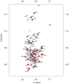
Figure 1
Figure 1. Residues 14–43 of DARC contain the minimal binding region.
1H-15N-TROSY spectra of unbound DARC 1–60 (black) overlaid on 1H-15N-TROSY spectra of DARC 1–60 in the presence of excess unlabelled DBP-RII (red). Sequence assignments are shown for the unbound DARC 1H-15N-TROSY spectra. Peaks still visible in the presence of DBP-RII (red) are at DARC 1–60's N- and C- termini. Residues that disappear in the presence of DBP-RII are in the center of DARC and delineate the binding region.
Figure 2
Figure 2. Crystal Structure of the DBP-RII∶DARC heterotrimer and heterotetramer.
Overview of (A) DBP-RII∶DARC heterotrimer and (B) the DBP-RII∶DARC heterotetramer. Rotated views, (C) and (D), show DARC helices are oriented in parallel in the heterotetramer. DBP-RII monomers are in yellow and green. DARC monomers are in purple and blue.
Figure 3
Figure 3. Isothermal titration calorimetry reveals step-wise binding of DARC to DBP-RII in solution.
(A) A biphasic binding profile is observed indicating the formation of the heterotrimer at a molar ratio of 0.5 (n1 = 0.44±0.02, Kd1 = 2183±125 nM, ΔH1 = −2663±69 cal/mol) and heterotetramer at a molar ratio of 1 (n2 = 0.50±0.02, Kd2 = 88.5±26.6 nM, ΔH2 = −3338±23 cal/mol). The fit to the two independent site binding model is shown as a red line. Molar ratios are expressed as monomers of DBP-RII. Open circles denote unbound DBP, closed circles denote bound DBP. Titration of (B) PBS into DBP and (C) DARC into PBS reveals no observable profiles demonstrating the biphasic profile is due to DARC binding to DBP. In all cases, the top panel contains raw binding data, and the bottom panel changes in enthalpy associated with binding.
Figure 4
Figure 4. Binding interfaces of the DBP-RII∶DARC heterotrimer.
(A) Global view of the DBP-RII∶DARC heterotrimer, showing (B) DARC monomer A interactions and (C) the DBP-RII homodimeric interface. DARC monomer A is in purple, DBP-RII monomer 1 is in green and DBP-RII monomer 2 is in yellow. Residue numbers are labeled and DARC residue labels are underlined.
Figure 5
Figure 5. Binding interfaces of the DBP-RII∶DARC heterotetramer.
(A) Global view of the DBP-RII∶DARC heterotetramer, showing (B) the DBP-RII homodimeric interface, (C) DARC monomer A interactions, and (D) DARC monomer B interactions. DARC monomer A is in purple, DARC monomer B is in blue, DBP-RII monomer 1 is in green and DBP-RII monomer 2 is in yellow. Residue numbers are labeled and DARC residue labels are underlined.
Figure 6
Figure 6. The structural studies define red blood cell binding.
(A) Adherent HEK293 cells in grey bind to darker, smaller red blood cells when transfected with a DBP-RII surface expression plasmid with a GFP marker. Red blood cell rosetting images for DBP-RII mutants, showing bright field (left), GFP (center), and merged images (right). (B) Percentage of cells expressing point mutants which bind red blood cells, relative to wildtype, shown with standard error. (C) The major DBP-RII∶DARC residues identified in the crystal structures are indicated by red dots. Non-conservative P. knowlesi mutations at critical DBP-RII∶DARC contact residues 274, 356, and 363 suggest why PkDBPα but not PkDBPβ or PkDBPγ bind DARC. (D) Red blood cell rosetting images for DBP-RII receptor specificity mutants, showing bright field (left), GFP (center), and merged images (right). (E) Percentage of cells expressing receptor specificity point mutants which bind red blood cells, relative to wildtype, shown with standard error.
Figure 7
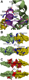
Figure 7. Mapping polymorphic residues and inhibitory epitopes reveals targets of selective pressure.
DBP-RII molecules are in green and yellow. DARC molecules are in purple and blue. DARC residue labels are underlined. (A) Nonsynonymous DARC polymorphisms in primates, residues colored in blue, which make critical contacts with DBP-RII provide a mechanism for inter-species transmission barriers. (B) Polymorphic DBP residues, in blue, are spread throughout the molecule. The most polymorphic region of DBP is the “DEK epitope” opposite the DARC14–43 binding site. (C) Inhibitory epitopes, in red and brown, map to the heterotetramer interface, DARC binding pockets and RBC proximal face of DBP-RII.
Figure 8
Figure 8. A model for attachment during invasion.
An initial binding event is followed by receptor-induced dimerization, as in the DBP-RII∶DARC heterotrimer. This brings a second DBP-RII molecule in close proximity to a second DARC ectodomain in the DARC homodimer. A second binding event creates the DBP-RII∶DARC heterotetramer. DBP-RII molecules are in green and yellow and DARC19–30 molecules are in purple and blue. The DARC homodimer is represented by a homology model. A schematic for the stepwise assembly is shown at the bottom. Closed circle – bound DBP-RII, open circle – unbound DBP-RII.

References

    1. Mendis K, Sina BJ, Marchesini P, Carter R (2001) The neglected burden of Plasmodium vivax malaria. Am J Trop Med Hyg 64: 97–106. - PubMed
    1. Price RN, Tjitra E, Guerra CA, Yeung S, White NJ, et al. (2007) Vivax malaria: neglected and not benign. Am J Trop Med Hyg 77: 79–87. - PMC - PubMed
    1. Welch SG, McGregor IA, Williams K (1977) The Duffy blood group and malaria prevalence in Gambian West Africans. Trans R Soc Trop Med Hyg 71: 295–296. - PubMed
    1. Howes RE, Patil AP, Piel FB, Nyangiri OA, Kabaria CW, et al. (2011) The global distribution of the Duffy blood group. Nat Commun 2: 266. - PMC - PubMed
    1. Miller LH, Mason SJ, Clyde DF, McGinniss MH (1976) The resistance factor to Plasmodium vivax in blacks. The Duffy-blood-group genotype, FyFy. N Engl J Med 295: 302–304. - PubMed

Publication types

MeSH terms