. 2014 Jan;10(1):e1003969.
doi: 10.1371/journal.pgen.1003969. Epub 2014 Jan 9.

Deletion of a conserved cis-element in the Ifng locus highlights the role of acute histone acetylation in modulating inducible gene transcription

Affiliations

Deletion of a conserved cis-element in the Ifng locus highlights the role of acute histone acetylation in modulating inducible gene transcription

Anand Balasubramani et al. PLoS Genet. 2014 Jan.

Abstract

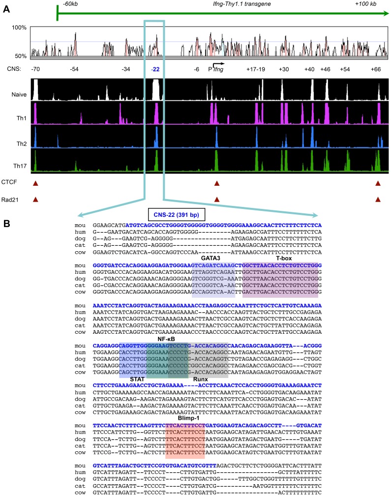
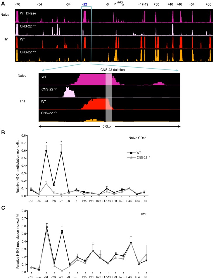
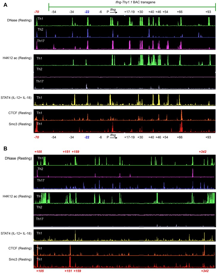
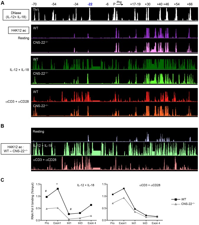
Differentiation-dependent regulation of the Ifng cytokine gene locus in T helper (Th) cells has emerged as an excellent model for functional study of distal elements that control lineage-specific gene expression. We previously identified a cis-regulatory element located 22 kb upstream of the Ifng gene (Conserved Non-coding Sequence -22, or CNS-22) that is a site for recruitment of the transcription factors T-bet, Runx3, NF-κB and STAT4, which act to regulate transcription of the Ifng gene in Th1 cells. Here, we report the generation of mice with a conditional deletion of CNS-22 that has enabled us to define the epigenetic and functional consequences of its absence. Deletion of CNS-22 led to a defect in induction of Ifng by the cytokines IL-12 and IL-18, with a more modest effect on induction via T-cell receptor activation. To better understand how CNS-22 and other Ifng CNSs regulated Ifng transcription in response to these distinct stimuli, we examined activation-dependent changes in epigenetic modifications across the extended Ifng locus in CNS-22-deficient T cells. We demonstrate that in response to both cytokine and TCR driven activation signals, CNS-22 and other Ifng CNSs recruit increased activity of histone acetyl transferases (HATs) that transiently enhance levels of histones H3 and H4 acetylation across the extended Ifng locus. We also demonstrate that activation-responsive increases in histone acetylation levels are directly linked to the ability of Ifng CNSs to acutely enhance Pol II recruitment to the Ifng promoter. Finally, we show that impairment in IL-12+IL-18 dependent induction of Ifng stems from the importance of CNS-22 in coordinating locus-wide levels of histone acetylation in response to these cytokines. These findings identify a role for acute histone acetylation in the enhancer function of distal conserved cis-elements that regulate of Ifng gene expression.


Conflict of interest statement

The authors have declared that no competing interests exist.

Figures

Figure 1
Figure 1. Ifng-CNS-22: An evolutionarily conserved distal regulatory hub.
(A) Syntenic regions of human and murine Ifng gene loci are shown aligned using the VISTA browser. CNSs represent regions of at least 100 bp in length that exhibit at least 70% homology between these two species. Locations of key Ifng CNSs including CNS-22 are listed below. Aligned below are DNase I hypersensitivity tracks of the extended Ifng locus generated from naïve CD4+, Th1, Th2 and Th17 cells. DNase I tracks represent averages from two independent runs. CTCF and cohesion binding sites that represent putative boundaries of the Ifng locus are highlighted below. Also shown is the extent of the Ifng-Thy1.1 (green) BAC transgene that we previously employed to study cis regulation of IFN-γ. This transgene spanned from −60 kb upstream to +100 kb downstream of Ifng, but excluded a more recently identified insulator sequence located 70 kb upstream of Ifng. (B) Genomic sequence of region deleted to generate CNS-22−/− mice is shown aligned against syntenic sequences from the genomes of multiple mammalian species. The murine CNS-22 sequence deleted to generate CNS-22−/− mice is highlighted in blue. The same sequence highlighted in this figure was also deleted from the Ifng-Thy1.1 transgene in our previous studies. Also highlighted are trans factor binding sites of transcription factors that have been demonstrated to bind to CNS-22.
Figure 2
Figure 2. IL-12+IL-18 driven Ifng transcription is compromised in CNS-22−/− T cells and NK cells.
(A) CD4+ T cells derived from OT-II transgenic WT and CNS-22−/− mice were differentiated with 2 ng/ml IL-12, 5 µg/ml ova-peptide (ISQAVHAAHAEINEAGR) and cultured with CD4-depleted irradiated feeder cells derived from Il12a −/− mice. Cells were reactivated for 4 h as described in methods and expression of IFN-γ was assessed by flow cytometric analysis following intracellular staining. Percentages of viable, IFN-γ+ T cells are indicated by black numbers and mean fluorescence intensities (MFI) of IFN-γ+ cells are indicated in grey. Data are representative of at least three independent experiments. (B) CD8+ T cells isolated from WT and CNS-22−/− mice were differentiated with 2.5 µg/ml of anti-CD3 antibody, 2 ng/ml IL-12 for 3 days and reactivated for 4 h with IL-12+IL-18 and subject to intracellular staining. For evaluating IFN-γ expression in NK cells, following depletion of both CD4+ and CD8+ T cells bulk splenocytes were activated with IL-12+IL-18 for 4 h and subject to intracellular staining. Percentages of viable, IFN-γ+ T/NK cells are indicated in black and MFI of IFN-γ+ cells are indicated in grey. Data are representative of at least three independent experiments. (C) Total RNA from anti-CD3+anti-CD28 or IL-12+IL-18 stimulated WT and CNS-22−/− Th1 cells was isolated at indicated time points and reverse-transcribed to generate cDNA. Transcript levels were measured by RT-PCR and relative levels of spliced transcripts were calculated by normalization against levels of spliced transcripts in resting Th1 cells. Data represent means from at least three independent experiments. Statistical analyses were carried out on means and standard errors from three independent experiments * p<0.01, # p<0.05, stimulated WT versus CNS-22−/−.
Figure 3
Figure 3. CNS-22 plays a limited role in long-range remodeling of the extended Ifng locus.
(A) Naïve CD4+ T cells and Th1 cells derived from WT and CNS-22−/− mice were subject to DNase-chip as previously described DNase I HS sites across the Ifng locus were analyzed with ACME using peak-calling thresholds set to a confidence limit of 95% and visualized with the IGB browser . DNaseI sites are shown aligned against a VISTA plot that highlights locations of Ifng CNSs. Data are representative of at least two independent experiments. (B, C) Naïve CD4+ T cells and Th1 cells derived from WT and CNS-22−/− mice were analyzed by ChIP using an antibody that recognizes all three methylation states of H3K4. Relative H3K4 methylation levels were calculated by comparisons with no antibody controls and are represented as a fraction of the H3K4 methylation observed at 16S ribosomal protein (16Srp) promoter, which was assigned a value of 1. Data represent mean±SEM from at least three independent experiments. * p<0.01, # p<0.05, stimulated WT versus CNS-22−/−.
Figure 4
Figure 4. Identification of putative regulatory elements greater than 100Ifng gene.
(A–B) Naïve CD4+ T cells from OT-II transgenic WT mice were differentiated under Th1, Th2 or Th17 polarizing conditions. Cells were left unstimulated or activated with IL-12+IL-18 for 1.5 h and subject to ChIP-chip. To identify STAT4, Smc3 and CTCF binding sites and to define H4K12ac-enriched regions across the Ifng locus, peak calling was carried out using a previously described algorithm for capturing microarray enrichment (ACME) . Peak-calling thresholds were set to a confidence limit of 95% and visualized with the IGB browser . Data are representative of at least two independent experiments.
Figure 5
Figure 5. Deletion of CNS-22 leads to prominent defects in IL-12+IL-18 dependent modulation of histone hyperacetylation.
(A) Th1 cells generated from either WT or CNS-22−/− mice were either left unstimulated or activated with IL-12+IL-18 for 1.5 h or anti-CD3 and anti-CD28 for 3 h and then subject to ChIP-chip with antibodies against H4K12ac. Levels of H4K12ac were analyzed as described in Fig. 3 and visualized using the IGB browser. (B) H4K12ac levels across the Ifng locus documented in WT Th1 cells were normalized against H4K12ac levels documented in CNS-22−/− Th1 cells to visualize in a semi-quantitative manner the magnitude to which locus-wide acquisition of H4K12ac marks was impaired in the absence of CNS-22. (C) Th1 cells generated from WT or CNS-22−/− mice were restimulated with anti-CD3+anti-CD28 for 3 h or IL-12+IL-18 for 1.5 h and recruitment of RNA-Pol II to the Ifng gene was assessed by ChIP-qPCR. RNA-Pol II recruitment is shown as a percentage of input DNA. Data represent means from at least three independent experiments. Statistical analyses were carried out on means and standard errors from three independent experiments * p<0.01, # p<0.05, stimulated WT versus CNS-22−/−.

References

    1. Ong CT, Corces VG (2011) Enhancer function: new insights into the regulation of tissue-specific gene expression. Nat Rev Genet 12: 283–293. - PMC - PubMed
    1. Frazer KA, Pachter L, Poliakov A, Rubin EM, Dubchak I (2004) VISTA: computational tools for comparative genomics. Nucleic Acids Res 32: W273–279. - PMC - PubMed
    1. Loots GG, Ovcharenko I, Pachter L, Dubchak I, Rubin EM (2002) rVista for comparative sequence-based discovery of functional transcription factor binding sites. Genome Res 12: 832–839. - PMC - PubMed
    1. Boyle AP, Davis S, Shulha HP, Meltzer P, Margulies EH, et al. (2008) High-resolution mapping and characterization of open chromatin across the genome. Cell 132: 311–322. - PMC - PubMed
    1. Crawford GE, Holt IE, Whittle J, Webb BD, Tai D, et al. (2006) Genome-wide mapping of DNase hypersensitive sites using massively parallel signature sequencing (MPSS). Genome Res 16: 123–131. - PMC - PubMed

Publication types

LinkOut - more resources