. 2014 Jan;10(1):e1004087.
doi: 10.1371/journal.pgen.1004087. Epub 2014 Jan 9.

Notch controls cell adhesion in the Drosophila eye

Affiliations

Notch controls cell adhesion in the Drosophila eye

Sujin Bao. PLoS Genet. 2014 Jan.

Abstract

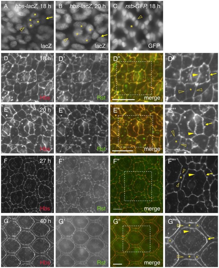
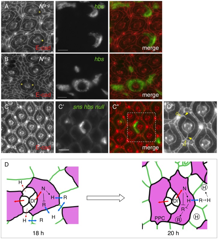
Sporadic evidence suggests Notch is involved in cell adhesion. However, the underlying mechanism is unknown. Here I have investigated an epithelial remodeling process in the Drosophila eye in which two primary pigment cells (PPCs) with a characteristic 'kidney' shape enwrap and eventually isolate a group of cone cells from inter-ommatidial cells (IOCs). This paper shows that in the developing Drosophila eye the ligand Delta was transcribed in cone cells and Notch was activated in the adjacent PPC precursors. In the absence of Notch, emerging PPCs failed to enwrap cone cells, and hibris (hbs) and sns, two genes coding for adhesion molecules of the Nephrin group that mediate preferential adhesion, were not transcribed in PPC precursors. Conversely, activation of Notch in single IOCs led to ectopic expression of hbs and sns. By contrast, in a single IOC that normally transcribes rst, a gene coding for an adhesion molecule of the Neph1 group that binds Hbs and Sns, activation of Notch led to a loss of rst transcription. In addition, in a Notch mutant where two emerging PPCs failed to enwrap cone cells, expression of hbs in PPC precursors restored the ability of these cells to surround cone cells. Further, expression of hbs or rst in a single rst- or hbs-expressing cell, respectively, led to removal of the counterpart from the membrane within the same cell through cis-interaction and forced expression of Rst in all hbs-expressing PPCs strongly disrupted the remodeling process. Finally, a loss of both hbs and sns in single PPC precursors led to constriction of the apical surface that compromised the 'kidney' shape of PPCs. Taken together, these results indicate that cone cells utilize Notch signaling to instruct neighboring PPC precursors to surround them and Notch controls the remodeling process by differentially regulating four adhesion genes.


Conflict of interest statement

The author has declared that no competing interests exist.

Figures

Figure 1
Figure 1. Notch is required for development of primary pigment cells (PPCs).
Eyes were stained using an anti-E-cadherin antibody in this figure. A–C) Wild type eyes at 18 h (A), 20 h (B) and 40 h (C) are shown. The tracings of eyes are shown in A′–C′, where PPC or PPCs precursors are highlighted in pink. D) Knockdown of Notch using a Notch RNAi transgene led to a failure of PPCs to develop. Typically, 3–5 cells (asterisks) were found adjacent to a cone cell cluster at 40 h APF. E) In the Nfa-g mutant, PPCs failed to develop. Frequently, 3–5 cells (asterisks) were contacting cone cells at 40 h APF. Scale bars, 10 µm.
Figure 2
Figure 2. Notch is activated in PPC precursors.
A) The Delta (Dl) protein was detected in cone cells (arrows) at 18 h APF as assessed with an anti-Dl antibody. B–C) Expression of a Dl reporter at 18 h (B) and 24 h APF (C) confirms its cone cell specificity. LacZ was detected at the highest level in anterior cone cells (arrows). D–E) Expression of GBE-Su(H)m8-lacZ, a reporter for Notch activity at 18 h (D) and 24 h APF (E). LacZ staining is shown on the left panel and E-cad channel in the middle. Merged views are shown on the right. Notch activity was high in cells adjacent to the anterior cone cells at 18 h APF (arrows, D). The difference of Notch activity between PPC precursors became less obvious at 24 h APF than earlier stages (arrows, E). Scale bars, 10 µm.
Figure 3
Figure 3. Notch signaling activates hbs and sns expression.
A–D) NICD or Dl (green) was over-expressed in single cells marked by GFP. The eyes were stained with an anti-Rst (left, A, C and D) or anti-lacZ antibody (left, B). Merged views are shown on the right. A) Over-expression of NICD increased Rst on the membrane. Arrows point to a PPC-IOC border with elevated Rst. B) Ectopic NICD induced ectopic hbs transcription as assessed using a hbs reporter (hbs-lacZ). Upon expression of NICD in a single IOC, ectopic lacZ was observed (arrow). C) When Dl was over-expressed in a posterior cone cell, ectopic Rst was detected at the border between the posterior PPC and its neighboring IOCs (arrow). D) When Dl was over-expressed in a polar cone cell, ectopic Rst was found at all borders surrounding the two PPCs (arrows). E–E″′) Notch is required for hbs transcription. In the Nfa-g mutant, hbs transcription (E) as assessed by the hbs reporter activity, was only detected in cone cells (bullets, E″) but lost in PPCs. In the Notch mutant, when Notch was activated in single IOCs by expressing NICD (E′), hbs transcription (arrows) as well as the characteristic ‘kidney’ shape of PPCs (asterisks) was restored. The lacZ and NICD channels are shown in E and E′, respectively, and the merged view in E″. The E-cadherin channel is shown in E″′. F–F″) Notch is required for sns expression. In the Nfa-g mutant, the Sns protein (F) was lost in PPCs. Sns expression in bristle groups (arrows) was not affected. In this mutant, when Notch was activated in single IOCs by expressing NICD (F′), the Sns protein (open arrowheads) was restored. The Sns channel is shown in F and the NICD channel in F′. The merged view is shown in F″. Scale bars, 10 µm.
Figure 4
Figure 4. Notch signaling suppresses rst and kirre expression.
A–A″′) NICD (A′) was over-expressed in a single IOC and the Rst protein (A) was assessed using an anti-Rst antibody. The merged view is shown in A″. The enlarged view of a boxed region in A″ is shown in A″′. For clarity, only Rst channel is shown in A″′. Rst was increased at IOC-IOC borders (arrowheads) but reduced at the PPC-IOC border (arrow). Rst was undetectable at wild type IOC-IOC borders (open arrowheads). Single IOCs (asterisks) and bristle groups (carets) are indicated. B–B″′) NICD (B′) was over-expressed in a single IOC and the rst transcript (B) was assessed using a rst reporter (rstF6-lacZ). Merged view is shown in B″ and cell shape was visualized using an anti-DE-cadherin antibody (B″′). Arrows point to IOCs that lost lacZ staining. C–C″) Notch is required to suppress rst transcription. In the Nfa-g mutant, the rst transcript was detected in cells adjacent to cone cells (open arrowheads). Cell morphology was visualized using an anti-Armadillo (Arm) antibody (C). rst transcription was assessed using the rst reporter rstF6-lacZ (C′). The merged view is shown in C″. D–D″) NICD (green, D′) was over-expressed in a single IOC and the eye was stained with an anti-Kirre antibody (D). The merged view is shown in D′. The enlarged view of a boxed region in D′ is shown in D″. For clarity, only Kirre channel is shown in D″. The Kirre protein was increased at an IOC-IOC border (arrowhead) but reduced at the PPC-IOC border (arrow). Kirre was undetectable at wild type IOC-IOC borders (open arrowheads). Single IOCs (asterisks) and bristle groups (carets) are indicated. Scale bars, 10 µm.
Figure 5
Figure 5. Distribution of Hbs and Rst is dynamic in the eye epithelium.
The activity of a hbs reporter was assessed using an anti-lacZ antibody (A–B). Cone cells are marked (asterisks). A) The activity of the hbs reporter was detected in emerging PPCs at 18 h APF. The nucleus of a PPC precursor (arrow) was rising half way to the level of cone cell nuclei while the nucleus of the second PPC precursor was lagging behind (open arrowhead). B) The activity of the hbs reporter was detected in PPC precursors at 20 h APF. The nucleus of a PPC precursor (arrow) had arisen to the level of those of cone cells while the nucleus of the second PPC precursor was still below the plane (open arrowhead). C) rst was transcribed in IOCs (open arrowhead) at 18 h APF. rst-Gal4 was used to drive expression of nuclear GFP(rst>GFP). The location for an ommatidium is indicated (asterisk). D–G″′) Distribution of Hbs and Rst is dynamic during 18–40 h APF. The Hbs channels (red) are shown in D–G and the Rst channels (green) in D′–G′. The merged views are shown on D″–G″. The enlarged views of boxed regions in D″–G″ are shown in D″′–G″′. For clarity, only Hbs channels are shown in D″′–G″′. Single IOCs (asterisks) and bristle groups (carets) are indicated. D) At 18 h APF, both Hbs and Rst were found at all borders surrounding PPC precursors including PPC-IOC borders (arrows) and PPC-cone borders (arrowheads). These proteins were also present at IOC-IOC borders (open arrowheads). E) At 20 h APF, PPCs fully enwrapped cone cells. Both Hbs and Rst were enriched at PPC-IOC (arrows), PPC-cone and PPC-PPC borders (arrowheads) while these proteins were slightly reduced at IOC-IOC borders (open arrowheads). F) At 27 h APF, both Hbs and Rst were enriched at PPC-IOC borders (arrows) while these proteins were reduced at PPC-PPC and PPC-cone borders (arrowheads). Hbs and Rst were diminished at IOC-IOC borders (open arrowheads). G) At 40 h APF, both Hbs and Rst proteins were enriched at IOC-PPC borders (arrows) while they were diminished at PPC-PPC and PPC-cone borders (arrowhead). Hbs and Rst were undetectable at IOC-IOC borders (open arrowheads). Scale bars, 10 µm.
Figure 6
Figure 6. cis-interactions promote protein turnover.
A–A″) Hbs promotes turnover of Rst in the same cell. hbs was mis-expressed in a single IOC (A′) and the eye was stained with an anti-Rst antibody (A). Levels of Rst were reduced at the border between the target IOC and its neighboring PPC (arrow) and increased in vesicles (open arrowhead). Merged view is shown in A′. The enlarged view of a boxed region in A′ with the Rst channel is shown in A″. B–B″) Rst promotes turnover of Hbs in the same cell. rst was mis-expressed in single cells (B′) and the eye stained with an anti-Hbs antibody (B). The Hbs level was reduced at PPC-IOC borders (arrows). A higher level of the Hbs protein was observed in vesicles (open arrowheads). When Rst was expressed in a cone cell, the Hbs level was elevated at the cone-PPC border (arrows). The target cone cell also had a higher level of Hbs in vesicles (open arrowheads). Merged view is shown in B′. The enlarged view of a boxed region in B′ with the Hbs channel is shown in B″. C) Interference of Hbs by mis-expressing Rst in PPCs (spa>rst) led to severe disruption of the hexagonal pattern of the eye. Three cells surrounding a cone cell cluster are highlighted (asterisks). IOCs failed to sort into a single file (open arrowheads). D) Over-expression of N-cadherin in cone cells (spa>N-cadherin) had a mild effect on tissue remodeling. An abnormal cone cell was highlighted (arrow) along with a defective IOC (open arrowhead). E) Over-expression of Rst in IOCs (54>rst) had a mild effect on tissue remodeling. Several IOCs formed a cluster around a bristle group (open arrowheads). Scale bars, 10 µm.
Figure 7
Figure 7. Spatial organization of primary pigment cells requires Hbs and Sns.
Eyes were stained with an anti-E-cadherin antibody (red, left). Target (mutant or over-expression) cells are marked by GFP (green, middle). The merged views are shown on the right. A–B) Expression of Hbs in a single cell restored the ‘kidney’ shape of PPCs. In the Nfa-g eye, hbs was expressed in single IOCs. When hbs was expressed in single cells adjacent to cone cells, the cell (asterisks) spread around cone cells (A). When hbs was introduced into two cells adjacent to cone cells, these cells (asterisks) fully enwrapped cone cells (B). C–C″′) Hbs and Sns are required for organization of PPCs. In a single PPC mutant for both hbs and sns (green, C′), the cell reduced the apical surface area and PPC-IOC border. The enlarged view of a boxed region in C″ with the E-cadherin channel is shown in C″′. Open arrowheads mark the shortened PPC-IOC border while arrowheads highlight the curved PPC-PPC borders. D) A model for control of PPC recruitment by cell signaling and cell adhesion. At 18 h APF, all IOCs that contact cone cells have access to Dl and express Hbs. However, IOCs adjacent to anterior-posterior cone cells receive a high level of Notch signaling (thick red lines) than other IOCs (thin red lines). These cells express a higher level of Hbs than other IOCs. Hbs boosts the ability of these cells to enwrap cone cells and gain more access to Dl. Therefore, Notch signaling and Hbs create a positive feedback loop so that initially a small difference in Notch signaling is amplified. As a result, two cells adjacent to anterior and posterior cone cells outcompete other IOCs and enwrap cone cells as PPC precursors. At 20 h APF, PPC precursors gain full access to Dl and constantly produce Hbs while shutting down Rst production. The remaining Rst in PPCs is removed by cis-interactions. In the meantime, other IOCs that are now denied access to Dl constantly supply Rst. The remaining Hbs in IOCs are cleared out by cis-interactions, leading to complementary distribution of two groups of adhesion molecules. For simplicity, only Hbs and Rst are shown. H = Hbs; R = Rst; N = Notch; Dl = Delta. Double-headed arrows represent trans-interactions between Hbs and Rst; Hbs and Rst in circle represent proteins in vesicles. Scale bars, 10 µm.

Similar articles

Cited by

References

    1. Barolo S, Posakony JW (2002) Three habits of highly effective signaling pathways: principles of transcriptional control by developmental cell signaling. Genes Dev 16: 1167–1181. - PubMed
    1. Dahmann C, Basler K (1999) Compartment boundaries: at the edge of development. Trends Genet 15: 320–326. - PubMed
    1. Irvine KD, Vogt TF (1997) Dorsal-ventral signaling in limb development. Curr Opin Cell Biol 9: 867–876. - PubMed
    1. Cagan RL, Ready DF (1989) Notch is required for successive cell decisions in the developing Drosophila retina. Genes Dev 3: 1099–1112. - PubMed
    1. Hoppe PE, Greenspan RJ (1986) Local function of the Notch gene for embryonic ectodermal pathway choice in Drosophila. Cell 46: 773–783. - PubMed

Publication types

MeSH terms

LinkOut - more resources