

. 2013 Nov 1:7:10.1038/nphoton.2013.284.
doi: 10.1038/nphoton.2013.284.
Recent Advances in Fiber Lasers for Nonlinear Microscopy
Affiliations
- PMID: 24416074
- PMCID: PMC3887125
- DOI: 10.1038/nphoton.2013.284
Item in Clipboard
Recent Advances in Fiber Lasers for Nonlinear Microscopy
Nat Photonics.
.
Abstract
Nonlinear microscopy techniques developed over the past two decades have provided dramatic new capabilities for biological imaging. The initial demonstrations of nonlinear microscopies coincided with the development of solid-state femtosecond lasers, which continue to dominate applications of nonlinear microscopy. Fiber lasers offer attractive features for biological and biomedical imaging, and recent advances are leading to high-performance sources with the potential for robust, inexpensive, integrated instruments. This article discusses recent advances, and identifies challenges and opportunities for fiber lasers in nonlinear bioimaging.
Figures
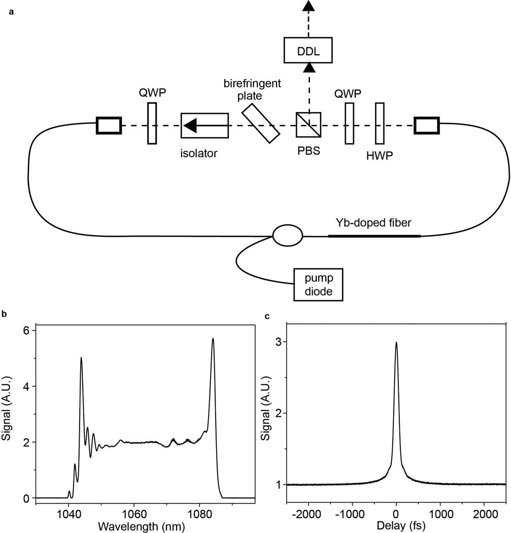
(a) Schematic of dissipative-soliton laser cavity. QWP: quarter wave plate, HWP: half wave plate, PBS: polarizing beam splitter, DDL: dispersive delay line. Spectrum (b) and autocorrelation (c) of 80-fs pulse from dissipative-soliton laser. From [40].
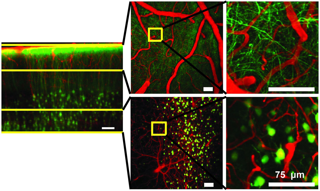
Images of the cortex of a live, anesthetized mouse with a glass-covered craniotomy, recorded in the laboratory of Prof. C. Schaffer at Cornell University. Pyramidal neurons are labeled by yellow fluorescent protein (green) and blood vessels are labeled by intravenously-injected Texas Red dextran (red). The top row shows surface vessels and dendritic processes in the top 150 µm; the bottom row shows arterioles, venules and capillaries as well as neuron cell bodies at depths of 450–650 µm. Yellow boxes (left) delineate areas of higher magnification (right). From Ref. .
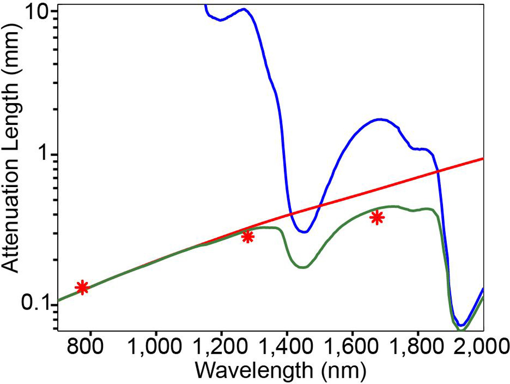
The water absorption length (blue line), the scattering length of mouse brain cortex (red line), and the effective attenuation length (green line). Note the logarithmic scale of the vertical axis. The scattering length is obtained by numerical simulation using Mie theory for a tissue-like phantom that resembles the scattering property of the cortex. The phantom contains 1 mm diameter polystyrene beads at a concentration of 5.4×10⁹/ml. The red stars indicate the reported attenuation lengths for mouse cortex in vivo from previous work. From Refs. and .
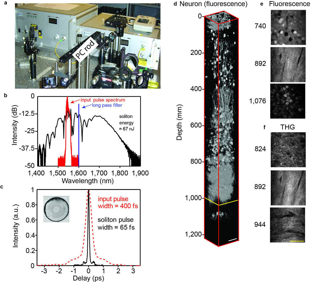
(a) Experimental setup of SSFS in a PC rod pumped by a fiber CPA system. The measured spectrum (b) and the corresponding second-order autocorrelation trace (c) of the input pulse (red) and the 1,675-nm soliton generated in the PC rod (black). The soliton energy, integrated from 1,617 nm, is 67 nJ. Inset in (c) shows the cross section of the PC rod. (d) 3D reconstruction of in vivo 3PM images in the brain of a mouse, which contains RFP-labelled pyramidal neurons. The external capsule extends from approximately 840 to 976 µm below the surface of the brain, and the stratum pyramidale extends from approximately 1,060 to 1,120 µm below the surface. Normalized x-y frames of the fluorescence (e) and THG (f) signal at various depths. The scale bar is 50 µm.
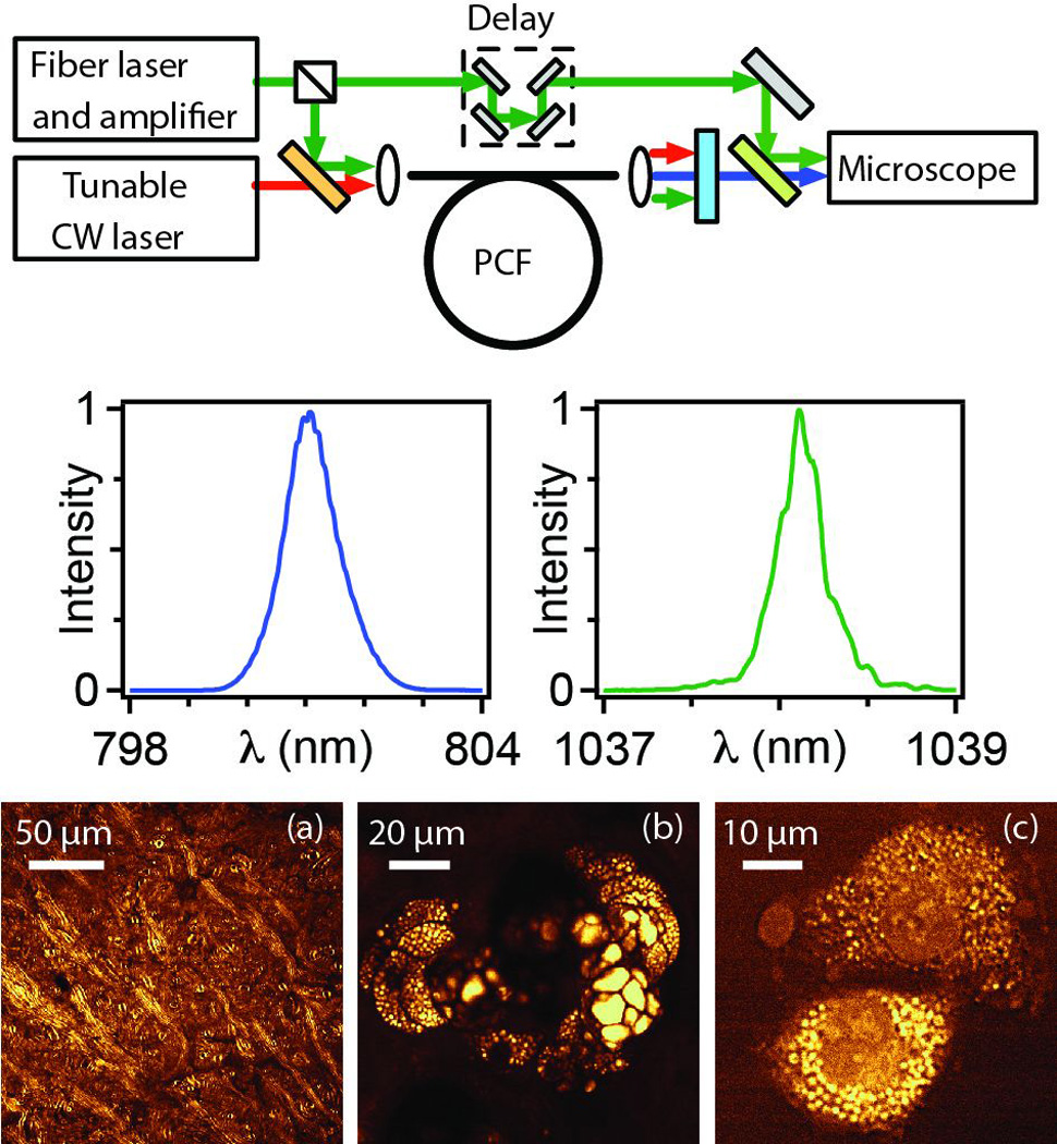
Top: schematic of fiber source based on FWM in PCF. Middle: spectra of generated pulses. Bottom: CARS images at the 2850 cm−1 mode of CH2: (a) mouse brain, (b) sebaceous gland 40 µm deep in mouse ear and (c) isolated rat fibroblast cells. From Ref. .
References
-
- Denk W, Strickler JH, Webb WW. Two-photon laser scanning fluorescence microscopy. Science. 1990;248:73–76. - PubMed
-
- Yuste R, Denk W. Dendritic spines as basic function units of neuronal integration. Nature. 1995;375:682–684. - PubMed
-
- Williams RM, Piston DW, Webb WW. Two-photon molecular excitation provides intrinsic 3-dimensional resolution for laser-based microscopy and microphotochemistry. FASEB J. 1994;8:804–813. - PubMed
-
- Denk W, Piston DW, Webb WW. Two-photon molecular excitation in laser scanning microscopy. In: Pawley J, editor. The Handbook of Confocal Microscopy. New York: Plenum; 1995. pp. 445–458.
-
- Helmchen F, Denk W. Deep tissue two-photon microscopy. Nat. Methods. 2005;2:932–940. - PubMed
Grants and funding
LinkOut - more resources
Full Text Sources
Other Literature Sources
Miscellaneous