. 2014 Jan 9;9(1):e84259.
doi: 10.1371/journal.pone.0084259. eCollection 2014.

Bridging the species divide: transgenic mice humanized for type-I interferon response

Affiliations

Bridging the species divide: transgenic mice humanized for type-I interferon response

Daniel Harari et al. PLoS One. .

Abstract

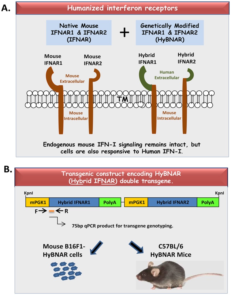
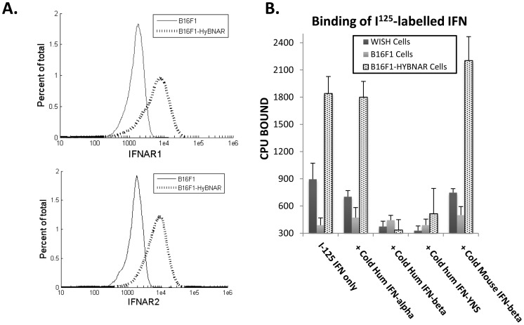
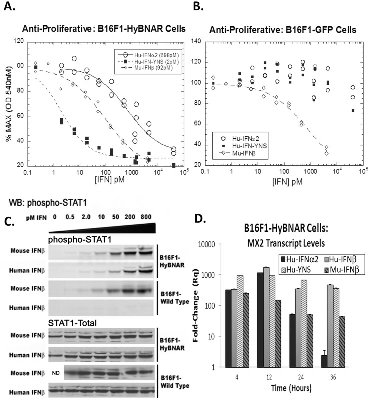
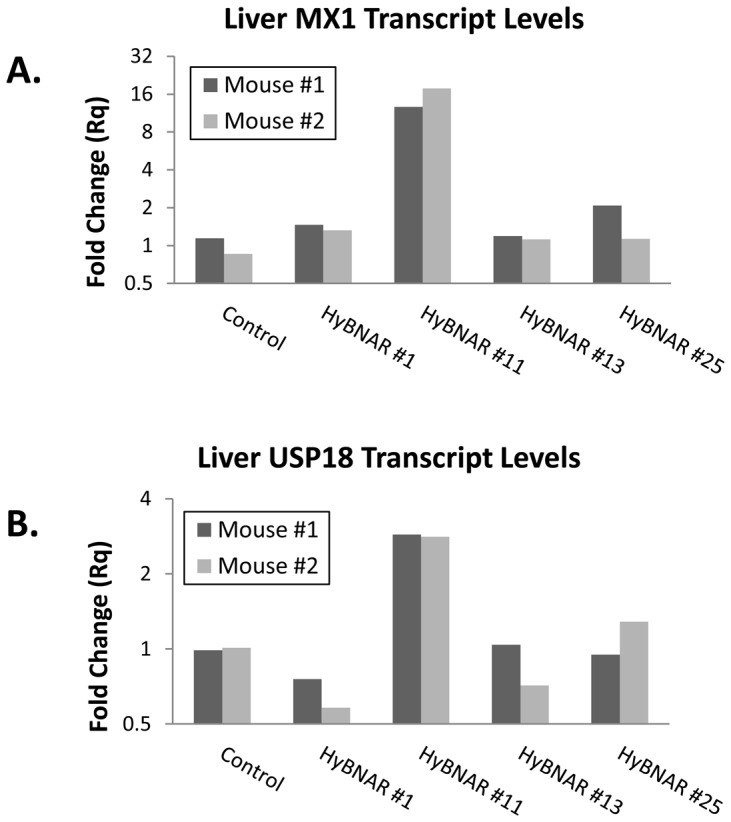
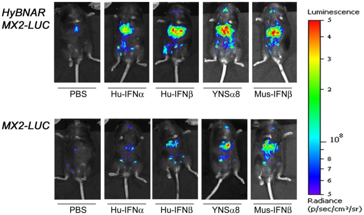
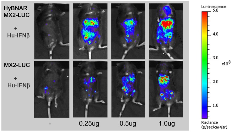
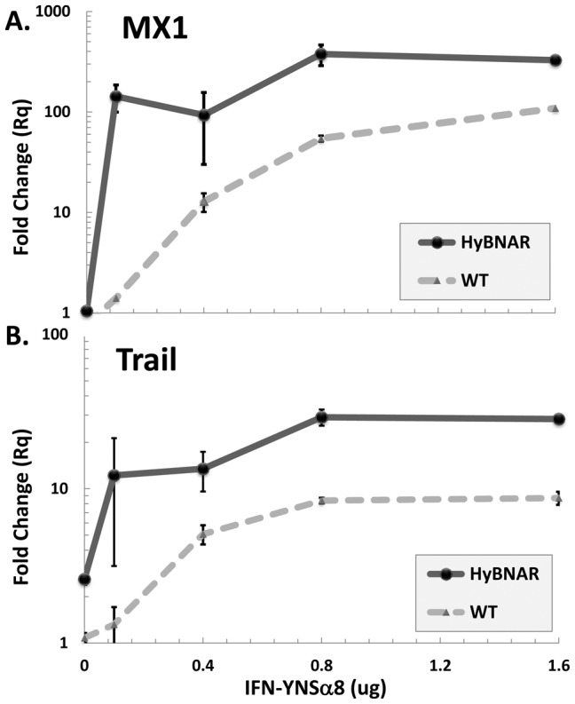
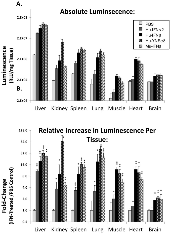
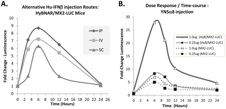
We have generated transgenic mice that harbor humanized type I interferon receptors (IFNARs) enabling the study of type I human interferons (Hu-IFN-Is) in mice. These "HyBNAR" (Hybrid IFNAR) mice encode transgenic variants of IFNAR1 and IFNAR2 with the human extracellular domains being fused to transmembrane and cytoplasmic segments of mouse sequence. B16F1 mouse melanoma cells harboring the HyBNAR construct specifically bound Hu-IFN-Is and were rendered sensitive to Hu-IFN-I stimulated anti-proliferation, STAT1 activation and activation of a prototypical IFN-I response gene (MX2). HyBNAR mice were crossed with a transgenic strain expressing the luciferase reporter gene under the control of the IFN-responsive MX2 promoter (MX2-Luciferase). Both the HyBNAR and HyBNAR/MX2-Luciferase mice were responsive to all Hu-IFN-Is tested, inclusive of IFNα2A, IFNβ, and a human superagonist termed YNSα8. The mice displayed dose-dependent pharmacodynamic responses to Hu-IFN-I injection, as assessed by measuring the expression of IFN-responsive genes. Our studies also demonstrated a weak activation of endogenous mouse interferon response, especially after high dose administration of Hu-IFNs. In sharp contrast to data published for humans, our pharmacodynamic readouts demonstrate a very short-lived IFN-I response in mice, which is not enhanced by sub-cutaneous (SC) injections in comparison to other administration routes. With algometric differences between humans and mice taken into account, the HyBNAR mice provides a convenient non-primate pre-clinical model to advance the study of human IFN-Is.


Conflict of interest statement

Competing Interests: A number of the authors of this manuscript are/were employed by Merck-Serono. Research performed was funded by Merck-KGaA (the parental company of Merck-Serono). Additionally, the authors declare that the disclosed patented technologies relating to IFN superagonists are associated with patent filings: USA: 60/694,810 and PCT/IL2006/000754 (along with downstream national filings). The authors will openly share their reagents described in this paper with academic institutions. This does not alter the authors' adherence to all the PLOS ONE policies on sharing data and materials.

Figures

Figure 1
Figure 1. Transgenic HyBNAR Mice and Cell Line.
IFN-Is transduce their signals to recipient cells via their binding to and transactivation of a heterodimeric cytoplasmic receptor complex containing IFNAR1 and IFNAR2. But human IFN-Is trans-activate the mouse IFNARs poorly. (A) To overcome this, we have introduced chimeric IFNAR receptors encoding the extracellular domains of IFNAR1 and IFNAR2, fused to their respective transmembrane and intracellular components of mouse origin. By using a regular transgeneisis methodology (IE: not by targeted IFNAR knock-in) we can ensure that regular mouse IFN-I signaling is not disrupted. (B) Diagram of the HyBNAR (Hybrid IFNAR) transgenic construct. Hybrid human and mouse IFNAR1 and IFNAR2 sequences were independently placed under the control of the weak but ubiquitously expressed mouse PGK1 promoter. The two transgenes were then linked together into a single construct. By stable transfection of this construct into the B16F1 mouse melanoma cell line or by pro-nuclear injection into the fertilized embryos of C57BL/6 donors, HyBNAR expressing cells and transgenic mice were generated.
Figure 2
Figure 2. Transgene expression on B16F1-HyBNAR mouse melanoma cells and its binding to human IFN-I.
(A) FACS analysis of B16F1-HyBNAR cells using antibodies that specifically bind to the human variants of the extracellular domains of IFNAR1 and IFNAR2. Specificity of binding is demonstrated by the staining of non-transfected mouse B16F1 control cells. (B) YNS, an engineered tight-binding variant of IFNα2 was radiolabelled and incubated with mouse B16F1, B16F1-HyBNAR and human WISH cells. Tightly binding radiolabelled YNS could not be displaced by co-incubation with 100× molar excess of lower affinity Hu-IFNα2a, although high affinity human IFNβ and YNS completely displaced the hot ligand. Importantly, 100× molar excess of mouse IFNβ did not displace binding of hot YNS in this system.
Figure 3
Figure 3. B16F1-HyBNAR mouse melanoma cells are sensitive to human IFN-Is.
(A & B) Mouse B16F1 melanoma cells were stably transfected with either the HyBNAR double transgenic construct, or a control GFP vector and tested for their anti-proliferative dose response to Hu-IFNα2, Hu-IFN-YNS and for comparison, mouse IFNβ. (A) HyBNAR transfected B16F1 cells were responsive to both human and mouse IFN-Is (EC50 values shown in parentheses) (B) In the GFP-transfected B16F1 control cells however, the human ligands only promoted a minor loss in proliferation and when administered at very high concentrations. (C) B16F1-HyBNAR and non-transfected control cells were incubated with an increasing dose of both human and mouse IFNβ. After 45 minutes incubation the cells were lysed and analyzed by Western Blot detection for phosphorylated STAT1. Total STAT1 measurements are shown as control. (D). B16F1-HyBNAR cells were treated for the indicated time-points with 100 pM human and mouse IFN-Is. Measurements of the IFN-response gene MX2 were performed by qPCR analysis. Relative fold-change was determined in comparison to untreated cells and normalized using the reference gene HPRT1. Error bars represent standard error of duplicate measurements.
Figure 4
Figure 4. Response of different HyBNAR mouse strains to human IFNβ stimulation.
Two representative mice from four independent transgenic HyBNAR strains were injected IV. with 400β. Six hours later, livers were collected and RNA was extracted for qPCR measurements. (A) Transcript levels for the IFN-response gene MX1 and (B) USP18. Relative fold-change was determined in comparison to untreated cells and normalized using the reference gene HPRT1.
Figure 5
Figure 5. Luciferase signal in HyBNAR/MX2-LUC and MX2-LUC mice in response to different IFN-Is.
Mice were injected IP with 1.0-Is. At T = 3.0 hours live animals were injected with luciferin, anaesthetized and live luminosity was measured by an image capturing (IVIS spectrum) device. For comparative purposes MX2-LUC mice without the HyBNAR transgene were also analyzed (bottom panel).
Figure 6
Figure 6. HyBNAR/MX2-LUC mice are both sensitive and responsive to human IFN-Is.
Transgenic mice expressing the luciferase reporter gene under the control of the MX2 promoter (MX2-LUC) were interbred with the HyBNAR mice. The HyBNAR/MX2-LUC mice (upper panel) were injected IP with increasing concentrations of human IFNβ. After 6 hours, mice were injected with luciferin, anaesthetized and live luminosity was measured by an image capturing device (IVIS spectrum). For comparative purposes MX2-LUC mice without the HyBNAR transgene were also analyzed (bottom panel).
Figure 7
Figure 7. HyBNAR homozygous mice are sensitized to human IFN-I response.
Dose response of homozygote HyBNAR after IP injection of increasing doses of the human superagonist IFN-YNSα8. After 6 hours, livers were collected and from the derived RNA/cDNA MX1 (A) and Trail (B) levels were determined by qPCR. Relative fold-change was determined in comparison to untreated wild-type (WT) mice and normalized using the reference gene HPRT1. The average of two mice were used for each injection dosage. The differences in dose response to IFN were significant when comparing control vs. HyBNAR mouse groups (p<0.0005 for both MX1 and Trail expression plots as determined by two-way paired Anova).
Figure 8
Figure 8. Luciferase activity measurements from tissue homogenates from HyBNAR/MX2-LUC mice.
Mice were injected IP with either PBS or 1.0 ug of the indicated type I Interferon. After six hours the mice were perfused with PBS and tissues were collected for homogenization and measured for luciferase activity. (A) Absolute values of luciferase activity standardized per unit wet weight for each tissue. (B) Relative up-regulation of luciferase signal per tissue, in relation to PBS injected controls. Significance values of IFN-treated samples in comparison to PBS controls are shown (one tailed T-Test; *: p≤0.05, **: p≤0.005).
Figure 9
Figure 9. Time and injection regiment affects response to IFN-Is.
(A). Response of different injection regiments to 0.2 ug (weight adjusted per 20 g mouse weight) of human IFNβ. Injections were by intraperitoneal (IP), intra venous (IV) or by sub-cutaneous (SC) route. (B). Mice were injected IP with the indicated increasing doses of YNSα8 and luminosity was measured in a time-course from live animal measurements. MX2-LUC mice doubly transgenic for HyBNAR (solid lines) or without presence of HyBNAR (dotted lines) were both measured. The relative luminosity values given are averaged from three animals (A) or two animals (B) per injection group and were measured from a defined region of maximal signal (found in the liver region) for each mouse as described in Figure S2. In both experiments, baseline luminosity measurements were determined from the mice taken immediately prior to IFN induction.
Figure 10
Figure 10. Interspecies Relatedness for a Selection of Cell Surface Receptors.
Interspecies protein sequences derived for IFNAR1, IFNAR2, the epidermal growth factor receptor (EGFR) and the Insulin Receptor (INSR) were each compared by respective pairwise BLASTP alignment. Percent amino acid identity scores are given. Pre-calculated percentage identity scores were extracted from the Homologene Server (NCBI, Release #67). Exceptionally, rat IFNAR2 protein sequence (accession #XP_001073550.1) was not available in the Homologene dataset, and was thus curated manually.

Similar articles

Cited by

References

    1. Borden EC, Sen GC, Uze G, Silverman RH, Ransohoff RM, et al. (2007) Interferons at age 50: past, current and future impact on biomedicine. Nat Rev Drug Discov 6: 975–990 10.1038/nrd2422 - DOI - PMC - PubMed
    1. Moraga I, Harari D, Schreiber G, Uzé G, Pellegrini S (2009) Receptor density is key to the alpha2/beta interferon differential activities. Mol Cell Biol 29: 4778–4787 10.1128/MCB.01808-08 - DOI - PMC - PubMed
    1. Levin D, Harari D, Schreiber G (2011) Stochastic receptor expression determines cell fate upon interferon treatment. Mol Cell Biol 31: 3252–3266 10.1128/MCB.05251-11 - DOI - PMC - PubMed
    1. Shen X, Hong F, Nguyen VA, Gao B (2000) IL-10 attenuates IFN-alpha-activated STAT1 in the liver: involvement of SOCS2 and SOCS3. FEBS Lett 480: 132–136. - PubMed
    1. Radaeva S, Jaruga B, Kim W-H, Heller T, Liang TJ, et al. (2004) Interferon-gamma inhibits interferon-alpha signalling in hepatic cells: evidence for the involvement of STAT1 induction and hyperexpression of STAT1 in chronic hepatitis C. Biochem J 379: 199–208 10.1042/BJ20031495 - DOI - PMC - PubMed

Publication types