

Identification of a novel Haemophilus parasuis-specific B cell epitope using monoclonal antibody against the OppA protein
- PMID: 24416241
- PMCID: PMC3887010
- DOI: 10.1371/journal.pone.0084516
Identification of a novel Haemophilus parasuis-specific B cell epitope using monoclonal antibody against the OppA protein
Abstract
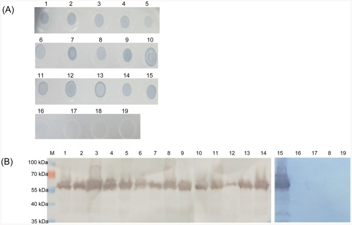
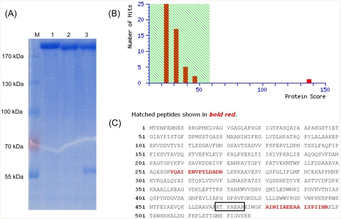
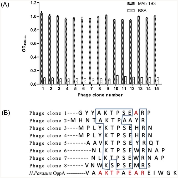
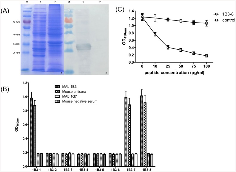
Monoclonal antibody (MAb) 1B3 against Haemophilus parasuis (H. parasuis) was generated by fusing SP2/0 murine myeloma cells and spleen cells from BALB/c mice immunized with the whole-bacterial-cell suspension of H. parasuis HS80 (serotype 5). The MAb 1B3 showed strong reactivity with 15 serotype reference strains of H. parasuis using Dot blot and Western blot analysis. Immunoprecipitation and protein spectral analysis indicated that MAb 1B3 recognized by Oligopeptide permease A (OppA) belongs to the ATP binding cassette transporter family. In addition, a linear B-cell epitope recognized by MAb 1B3 was identified by the screening of a phage-displayed 12-mer random peptide library. Sequence analysis showed that MAb 1B3 was recognized by phages-displaying peptides with the consensus motif KTPSEXR (X means variable amino acids). Its amino acid sequence matched (469)KTPAEAR(475) of H. parasuis OppA protein. A series of progressively truncated peptides were synthesized to define the minimal region that was required for MAb 1B3 binding. The epitope was highly conserved in OppA protein sequences from the isolated H. parasuis strains, which was confirmed by alignment analysis. Furthermore, the minimal linear epitope was highly specific among 75 different bacterial strains as shown in sequence alignments. These results indicated MAb 1B3 might be potentially used to develop serological diagnostic tools for H. parasuis.
Conflict of interest statement
Figures

Similar articles
-
Identification of a Novel Linear B-Cell Epitope of HbpA Protein from Glaesserella parasuis Using Monoclonal Antibody.Int J Mol Sci. 2023 May 12;24(10):8638. doi: 10.3390/ijms24108638. Int J Mol Sci. 2023. PMID: 37239984 Free PMC article.
-
Production and characterization of murine monoclonal antibodies against Haemophilus parasuis and study of their protective role in mice.Microbiology (Reading). 2004 Dec;150(Pt 12):3935-45. doi: 10.1099/mic.0.27443-0. Microbiology (Reading). 2004. PMID: 15583147
-
Immune response to oligopeptide permease A (OppA) protein in pigs naturally and experimentally infected with Haemophilus parasuis.Res Vet Sci. 2016 Aug;107:62-67. doi: 10.1016/j.rvsc.2016.05.006. Epub 2016 May 13. Res Vet Sci. 2016. PMID: 27473976
-
Identification of a conserved linear epitope on the VP1 protein of serotype O foot-and-mouth disease virus by neutralising monoclonal antibody 8E8.Virus Res. 2011 Jan;155(1):291-9. doi: 10.1016/j.virusres.2010.10.024. Epub 2010 Oct 23. Virus Res. 2011. PMID: 20974198
-
Identification of a conserved JEV serocomplex B-cell epitope by screening a phage-display peptide library with a mAb generated against West Nile virus capsid protein.Virol J. 2011 Mar 6;8:100. doi: 10.1186/1743-422X-8-100. Virol J. 2011. PMID: 21375771 Free PMC article.
Cited by
-
Identification of a Novel Linear B-Cell Epitope of HbpA Protein from Glaesserella parasuis Using Monoclonal Antibody.Int J Mol Sci. 2023 May 12;24(10):8638. doi: 10.3390/ijms24108638. Int J Mol Sci. 2023. PMID: 37239984 Free PMC article.
-
Identification of Two Novel Linear Neutralizing Epitopes within the Hexon Protein of Canine Adenovirus Using Monoclonal Antibodies.Vaccines (Basel). 2021 Feb 8;9(2):135. doi: 10.3390/vaccines9020135. Vaccines (Basel). 2021. PMID: 33567652 Free PMC article.
-
Evaluation of immunogenicity and protective efficacy of recombinant outer membrane proteins of Haemophilus parasuis serovar 5 in a murine model.PLoS One. 2017 Apr 27;12(4):e0176537. doi: 10.1371/journal.pone.0176537. eCollection 2017. PLoS One. 2017. PMID: 28448603 Free PMC article.
References
-
- Oliveira S, Pijoan C (2004) Haemophilus parasuis: new trends on diagnosis, epidemiology and control. Veterinary Microbiology 99: 1–12. - PubMed
-
- del Río ML, Martín CBG, Navas J, Gutiérrez-Muñiz B, Rodríguez-Barbosa JI, et al. (2006) aroA gene PCR-RFLP diversity patterns in Haemophilus parasuis and Actinobacillus species. Research in Veterinary Science 80: 55–61. - PubMed
-
- Rapp-Gabrielson VJ, Gabrielson DA (1992) Prevalence of Haemophilus parasuis serovars among isolates from swine. Am J Vet Res 53: 659–664. - PubMed
-
- Rafiee M, Bara M, Stephens CP, Blackall PJ (2000) Application of ERIC-PCR for the comparison of isolates of Haemophilus parasuis . Aust Vet J 78: 846–849. - PubMed
Publication types
MeSH terms
Substances
LinkOut - more resources
Full Text Sources
Other Literature Sources
Molecular Biology Databases
Miscellaneous
