

Specific relationship between excitatory inputs and climbing fiber receptive fields in deep cerebellar nuclear neurons
- PMID: 24416251
- PMCID: PMC3885585
- DOI: 10.1371/journal.pone.0084616
Specific relationship between excitatory inputs and climbing fiber receptive fields in deep cerebellar nuclear neurons
Abstract
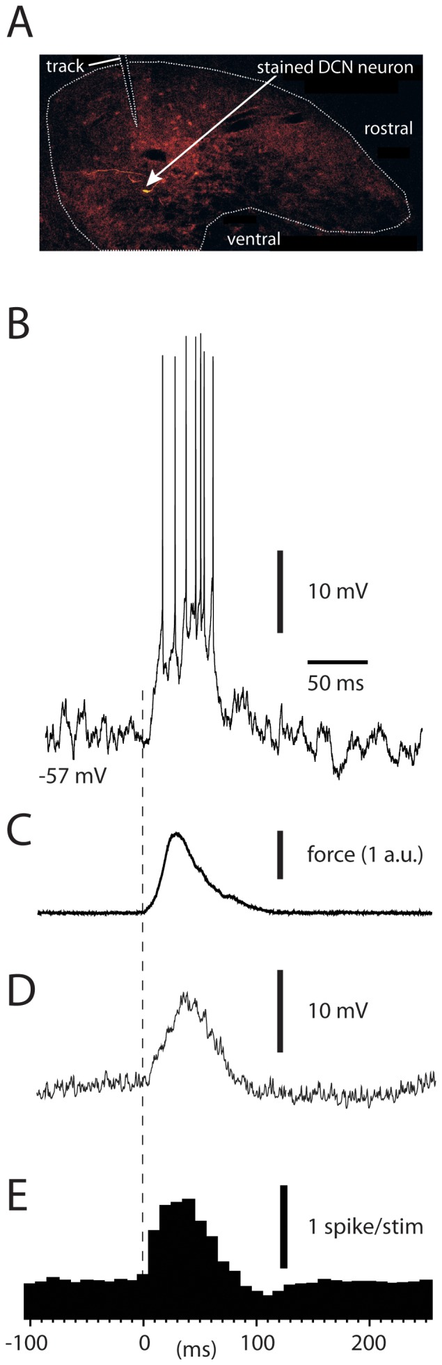
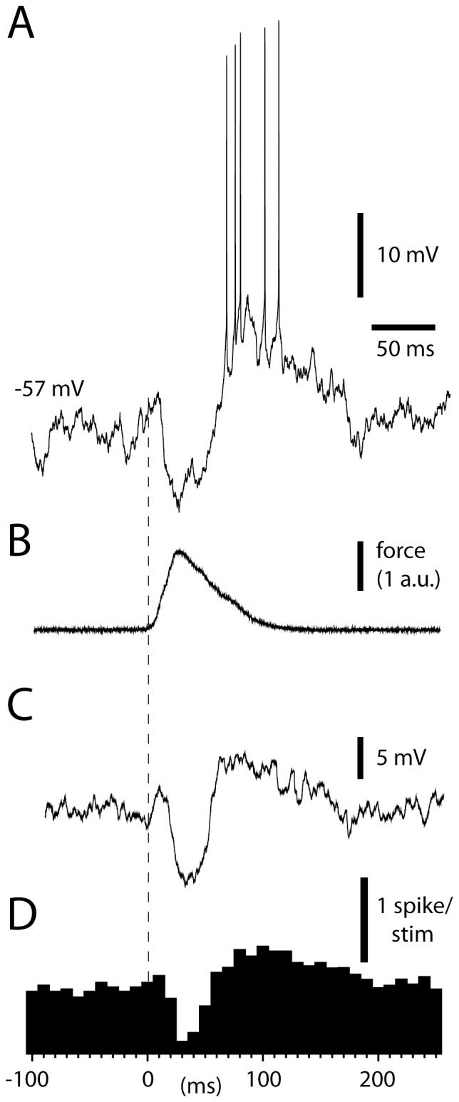
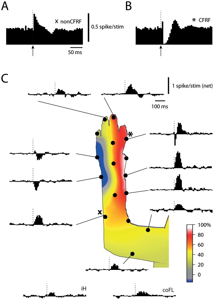
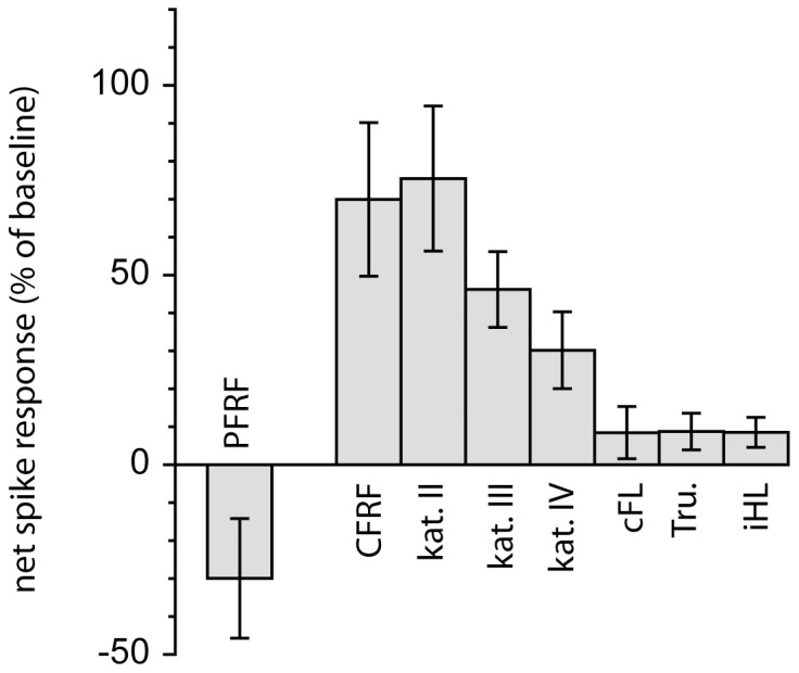
Many mossy fiber pathways to the neurons of the deep cerebellar nucleus (DCN) originate from the spinal motor circuitry. For cutaneously activated spinal neurons, the receptive field is a tag indicating the specific motor function the spinal neuron has. Similarly, the climbing fiber receptive field of the DCN neuron reflects the specific motor output function of the DCN neuron. To explore the relationship between the motor information the DCN neuron receives and the output it issues, we made patch clamp recordings of DCN cell responses to tactile skin stimulation in the forelimb region of the anterior interposed nucleus in vivo. The excitatory responses were organized according to a general principle, in which the DCN cell responses became stronger the closer the skin site was located to its climbing fiber receptive field. The findings represent a novel functional principle of cerebellar connectivity, with crucial importance for our understanding of the function of the cerebellum in movement coordination.
Conflict of interest statement
Figures
Similar articles
-
Climbing fiber receptive fields-organizational and functional aspects and relationship to limb coordination.Cerebellum. 2015 Jun;14(3):360-3. doi: 10.1007/s12311-015-0647-y. Cerebellum. 2015. PMID: 25598536 Review.
-
Functional relation between corticonuclear input and movements evoked on microstimulation in cerebellar nucleus interpositus anterior in the cat.Exp Brain Res. 1995;106(3):365-76. doi: 10.1007/BF00231060. Exp Brain Res. 1995. PMID: 8983981
-
Coding of tactile response properties in the rat deep cerebellar nuclei.J Neurophysiol. 2005 Aug;94(2):1236-51. doi: 10.1152/jn.00285.2005. J Neurophysiol. 2005. PMID: 16061491
-
Topographical organization of the cerebellar cortical projection to nucleus interpositus anterior in the cat.J Physiol. 1994 Jan 15;474(2):245-60. doi: 10.1113/jphysiol.1994.sp020017. J Physiol. 1994. PMID: 8006811 Free PMC article.
-
GABA-ergic transmission in deep cerebellar nuclei.Prog Neurobiol. 1997 Oct;53(2):259-71. doi: 10.1016/s0301-0082(97)00033-6. Prog Neurobiol. 1997. PMID: 9364613 Review.
Cited by
-
Widespread Decoding of Tactile Input Patterns Among Thalamic Neurons.Front Syst Neurosci. 2021 Feb 16;15:640085. doi: 10.3389/fnsys.2021.640085. eCollection 2021. Front Syst Neurosci. 2021. PMID: 33664654 Free PMC article.
-
Differential Coding Strategies in Glutamatergic and GABAergic Neurons in the Medial Cerebellar Nucleus.J Neurosci. 2020 Jan 2;40(1):159-170. doi: 10.1523/JNEUROSCI.0806-19.2019. Epub 2019 Nov 6. J Neurosci. 2020. PMID: 31694963 Free PMC article.
-
Climbing fiber receptive fields-organizational and functional aspects and relationship to limb coordination.Cerebellum. 2015 Jun;14(3):360-3. doi: 10.1007/s12311-015-0647-y. Cerebellum. 2015. PMID: 25598536 Review.
-
Cerebellar Modules and Their Role as Operational Cerebellar Processing Units: A Consensus paper [corrected].Cerebellum. 2018 Oct;17(5):654-682. doi: 10.1007/s12311-018-0952-3. Cerebellum. 2018. PMID: 29876802 Free PMC article. Review.
-
No Medium-Term Spinocerebellar Input Plasticity in Deep Cerebellar Nuclear Neurons In Vivo?Cerebellum. 2017 Jun;16(3):638-647. doi: 10.1007/s12311-016-0839-0. Cerebellum. 2017. PMID: 28032320 Free PMC article.
References
-
- Mason CR, Miller LE, Baker JF, Houk JC (1998) Organization of reaching and grasping movements in the primate cerebellar nuclei as revealed by focal muscimol inactivations. J Neurophysiol 79: 537–554. - PubMed
Publication types
MeSH terms
Grants and funding
LinkOut - more resources
Full Text Sources
Other Literature Sources
Miscellaneous
