

Contribution of the resting-state functional connectivity of the contralesional primary sensorimotor cortex to motor recovery after subcortical stroke
- PMID: 24416273
- PMCID: PMC3885617
- DOI: 10.1371/journal.pone.0084729
Contribution of the resting-state functional connectivity of the contralesional primary sensorimotor cortex to motor recovery after subcortical stroke
Abstract
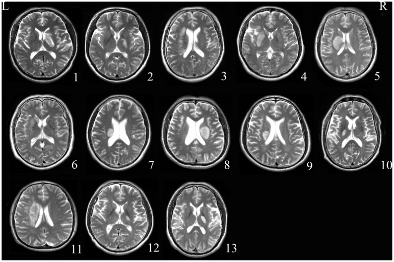
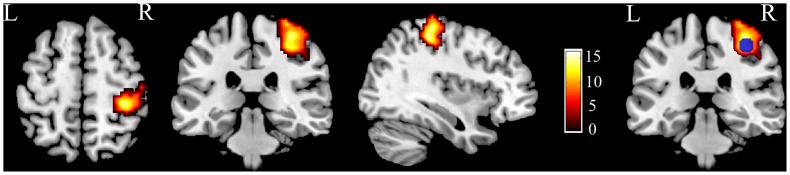
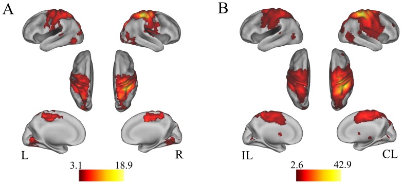
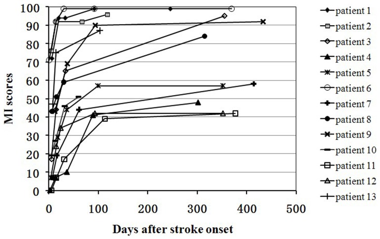
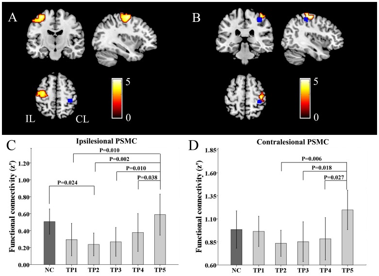
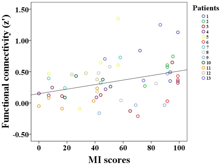
It remains uncertain if the contralesional primary sensorimotor cortex (CL_PSMC) contributes to motor recovery after stroke. Here we investigated longitudinal changes in the resting-state functional connectivity (rsFC) of the CL_PSMC and their association with motor recovery. Thirteen patients who had experienced subcortical stroke underwent a series of resting-state fMRI and clinical assessments over a period of 1 year at 5 time points, i.e., within the first week, at 2 weeks, 1 month, 3 months, and 1 year after stroke onset. Thirteen age- and gender-matched healthy subjects were recruited as controls. The CL_PSMC was defined as a region centered at the voxel that had greatest activation during hand motion task. The dynamic changes in the rsFCs of the CL_PSMC within the whole brain were evaluated and correlated with the Motricity Index (MI) scores. Compared with healthy controls, the rsFCs of the CL_PSMC with the bilateral PSMC were initially decreased, then gradually increased, and finally restored to the normal level 1 year later. Moreover, the dynamic change in the inter-hemispheric rsFC between the bilateral PSMC in these patients was positively correlated with the MI scores. However, the intra-hemispheric rsFC of the CL_PSMC was not correlated with the MI scores. This study shows dynamic changes in the rsFCs of the CL_PSMC after stroke and suggests that the increased inter-hemispheric rsFC between the bilateral PSMC may facilitate motor recovery in stroke patients. However, generalization of our findings is limited by the small sample size of our study and needs to be confirmed.
Conflict of interest statement
Figures
References
-
- Kwakkel G, Kollen B, Lindeman E (2004) Understanding the pattern of functional recovery after stroke: facts and theories. Restor Neurol Neurosci 22: 281–299. - PubMed
-
- Kwakkel G, Kollen B, Twisk J (2006) Impact of time on improvement of outcome after stroke. Stroke 37: 2348–2353. - PubMed
-
- Nudo RJ (2006) Mechanisms for recovery of motor function following cortical damage. Curr Opin Neurobiol 16: 638–644. - PubMed
-
- Cramer SC (2008) Repairing the human brain after stroke: I. Mechanisms of spontaneous recovery. Ann Neurol 63: 272–287. - PubMed
-
- Chollet F, DiPiero V, Wise RJ, Brooks DJ, Dolan RJ, et al. (1991) The functional anatomy of motor recovery after stroke in humans: a study with positron emission tomography. Ann Neurol 29: 63–71. - PubMed
Publication types
MeSH terms
LinkOut - more resources
Full Text Sources
Other Literature Sources
Medical
