

Initial imaging analysis of Budd-Chiari syndrome in Henan province of China: most cases have combined inferior vena cava and hepatic veins involvement
- PMID: 24416352
- PMCID: PMC3885682
- DOI: 10.1371/journal.pone.0085135
Initial imaging analysis of Budd-Chiari syndrome in Henan province of China: most cases have combined inferior vena cava and hepatic veins involvement
Abstract
Aim: To evaluate the type of venous involvement in Chinese Budd-Chiari syndrome (BCS) patients and the relative diagnostic accuracy of the different imaging modalities.
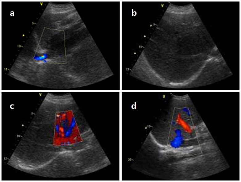
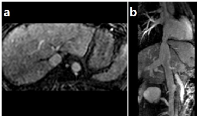
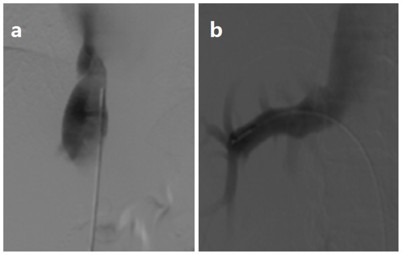
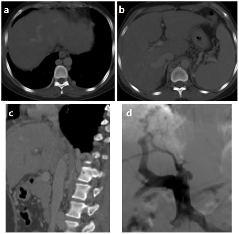
Methods: Using digital subtraction angiography (DSA) as a reference standard, color Doppler ultrasound (CDUS), computed tomography angiography (CTA), and magnetic resonance angiography (MRA) were performed on 338 patients with BCS. We analyzed the course of the main and any accessory hepatic veins (HVs) and the inferior vena cava (IVC) to assess the etiology of obstructed segments and diagnostic accuracy of CDUS, CTA and MRA.
Results: Among the 338 cases, there were 8 cases (2.4%) of isolated IVC membranous obstruction, 45 cases (13.3%) of isolated HV occlusion, and 285 cases (84.3%) with both IVC membranous obstruction and HV occlusion. Comparing with DSA, CDUS, CTA had a diagnostic accuracy of 89.3% and 80.2% in detecting BCS, and 83.4% of cases correctly correlated by MRA.
Conclusion: In Henan Province, most patients with BCS have complex lesions combining IVC and HV involvement. The combination of CDUS and CTA or MRI is useful for diagnosis of BCS and guiding therapy.
Conflict of interest statement
Figures
References
-
- Darwish Murad S, Valla DC, de Groen PC, Zeitoun G, Hopmans JA, et al. (2004) Determinants of survival and the effect of portosystemic shunting in patients with Budd-Chiari syndrome. Hepatology 39: 500–508. - PubMed
-
- Rav-Acha M, Gur C, Ilan Y, Verstandig A, Eid A (2004) [Budd-Chiari syndrome: updated treatment modalities]. Harefuah 143: 372–376, 389. - PubMed
-
- Okuda K (2002) Obliterative hepatocavopathy-inferior vena cava thrombosis at its hepatic portion. Hepatobiliary Pancreat Dis Int 1: 499–509. - PubMed
-
- Valla D, Hadengue A, el Younsi M, Azar N, Zeitoun G, et al. (1997) Hepatic venous outflow block caused by short-length hepatic vein stenoses. Hepatology 25: 814–819. - PubMed
-
- Plessier A, Rautou PE, Valla DC (2012) Management of hepatic vascular diseases. J Hepatol 56 Suppl 1S25–38. - PubMed
MeSH terms
LinkOut - more resources
Full Text Sources
Other Literature Sources
