. 2014 Jan 9;9(1):e85262.
doi: 10.1371/journal.pone.0085262. eCollection 2014.

SPBP is a sulforaphane induced transcriptional coactivator of NRF2 regulating expression of the autophagy receptor p62/SQSTM1

Affiliations

SPBP is a sulforaphane induced transcriptional coactivator of NRF2 regulating expression of the autophagy receptor p62/SQSTM1

Sagar Ramesh Darvekar et al. PLoS One. .

Abstract

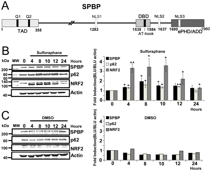
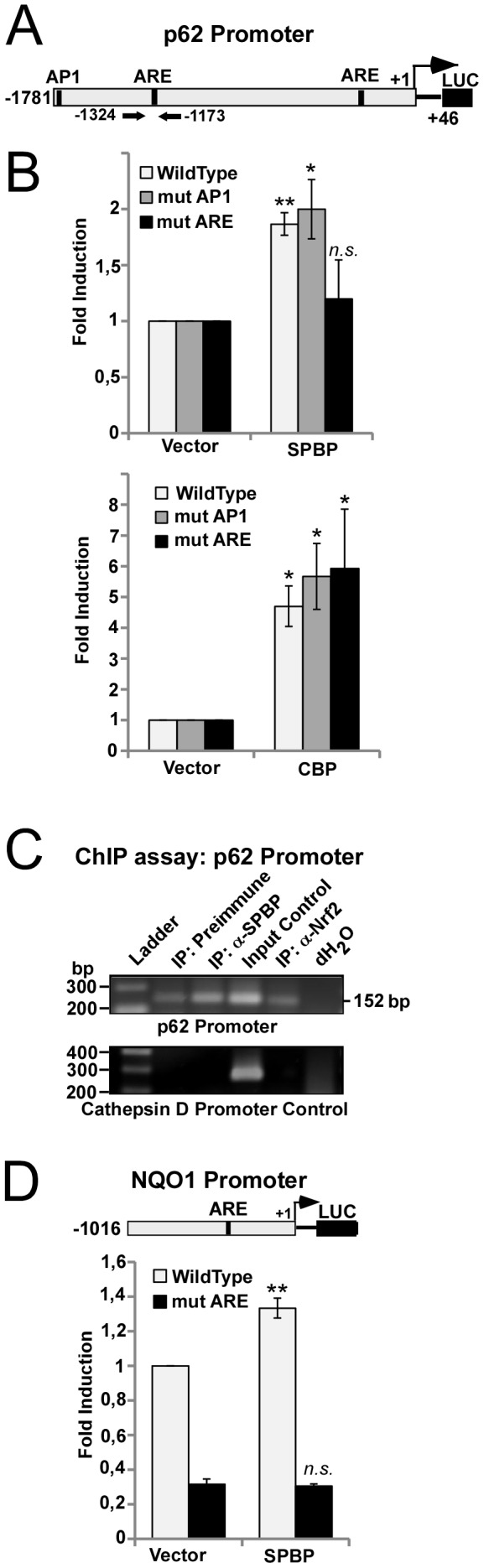
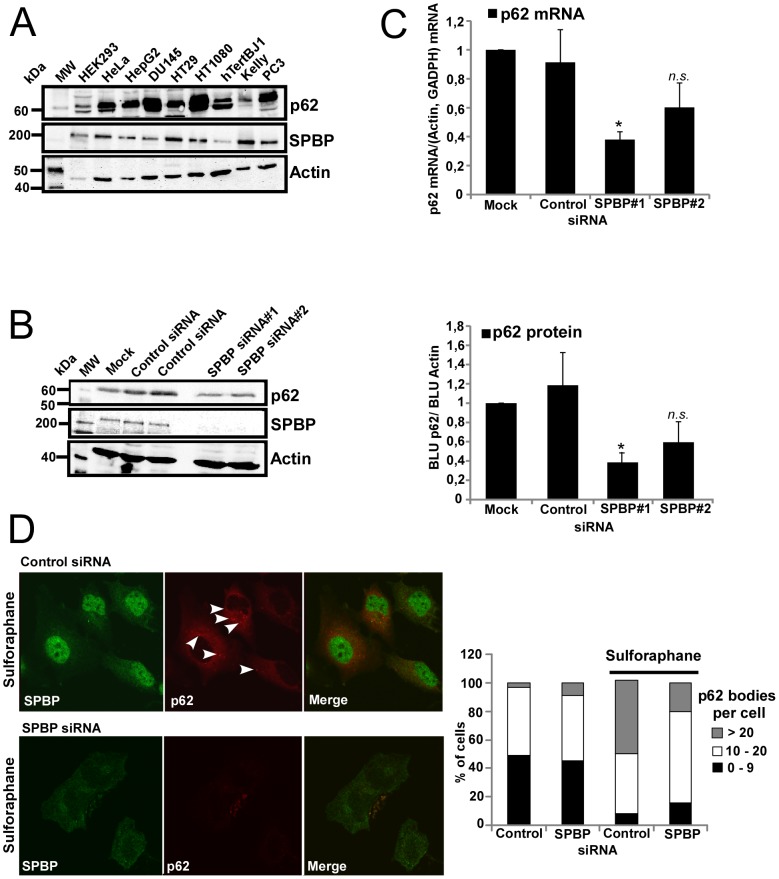
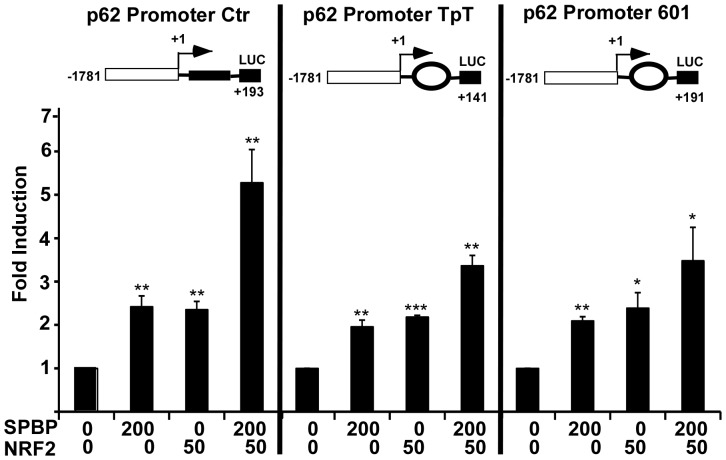
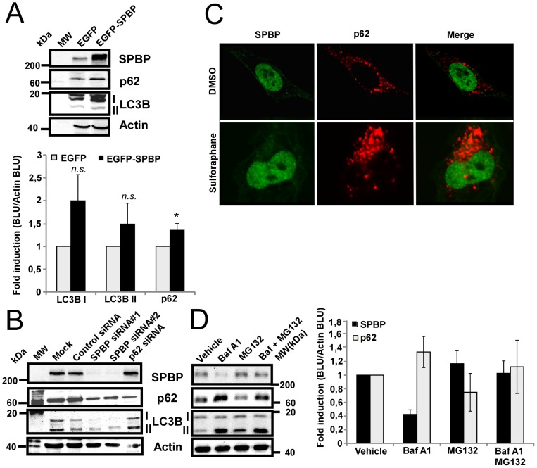
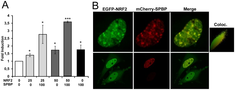
Organisms exposed to oxidative stress respond by orchestrating a stress response to prevent further damage. Intracellular levels of antioxidant agents increase, and damaged components are removed by autophagy induction. The KEAP1-NRF2 signaling pathway is the main pathway responsible for cell defense against oxidative stress and for maintaining the cellular redox balance at physiological levels. Sulforaphane, an isothiocyanate derived from cruciferous vegetables, is a potent inducer of KEAP1-NRF2 signaling and antioxidant response element driven gene expression. In this study, we show that sulforaphane enhances the expression of the transcriptional coregulator SPBP. The expression curve peaks 6-8 hours post stimulation, and parallels the sulforaphane-induced expression of NRF2 and the autophagy receptor protein p62/SQSTM1. Reporter gene assays show that SPBP stimulates the expression of p62/SQSTM1 via ARE elements in the promoter region, and siRNA mediated knock down of SPBP significantly decreases the expression of p62/SQSTM1 and the formation of p62/SQSTM1 bodies in HeLa cells. Furthermore, SPBP siRNA reduces the sulforaphane induced expression of NRF2, and the expression of the autophagy marker protein LC3B. Both these proteins contain ARE-like elements in their promoter regions. Over-expressed SPBP and NRF2 acts synergistically on the p62/SQSTM1 promoter and colocalize in nuclear speckles in HeLa cells. Collectively, these results suggest that SPBP is a coactivator of NRF2, and hence may be important for securing enhanced and sustained expression of NRF2 induced genes such as proteins involved in selective autophagy.


Conflict of interest statement

Competing Interests: The authors have declared that no competing interests exist.

Figures

Figure 1
Figure 1. Sulforaphane enhances SPBP expression.
(A) Schematic representation of the domain structure of human SPBP. TAD: trans-activation domain, DBD: DNA binding domain, NLS: Nuclear Localisation Signal, ePHD/ADD: extended PHD/ADD domain, Q1/Q2: Glutamine rich stretches. Numbers below indicate amino acid positions. (B) NRF2, SPBP and p62 display similar induction upon sulforaphane treatment. HeLa cells were exposed to 20 µM sulforaphane and cell extracts harvested for the indicated time points. Equivalent aliquots from the extracts were subjected to SDS-PAGE and western blot using specific anti-SPBP antibody, anti-p62 antibody, anti-NRF2 antibody or anti-actin as indicated. Fold induction calculated and correlated to actin in three independent experiments with standard deviations are shown to the right (**p<0.01, *p<0.05). (C) Control experiment showing that DMSO alone does not induce any changes in NRF2, SPBP or p62 expression levels. Equivalent aliquots of HeLa cell extracts exposed to DMSO and harvested for the indicated time points were subjected to SDS-PAGE and western blot using specific anti-SPBP antibody, anti-p62 antibody, anti-NRF2 antibody or anti-actin antibody as indicated. Fold induction calculated and correlated to actin are shown to the right.
Figure 2
Figure 2. SPBP mediates transcriptional activation via ARE elements.
(A) Schematic representation of the human −1781/+46 p62 promoter construct in front of the Luciferase gene. Conserved transcription factor binding sites relevant for this study are indicated. (B) Mutation of the ARE elements impairs SPBP mediated activation of the p62 promoter. Transient transfections were carried out in HEK293 cells using 60 ng of wild-type or mutated reporter vectors, and 100 ng of expression vectors for SPBP (upper panel) or CBP (lower panel). The data represent the mean of three independent experiments with standard deviations, each performed in triplicate (**p<0.01, *p<0.05, n.s. not significant). (C) Chromatin immunoprecipitations show that SPBP is associated with the p62 promoter. HeLa cell extracts (1.5×10⁷ cells per antibody) were immunoprecipitated with preimmune serum, polyclonal anti-SPBP antibody or polyclonal anti-NRF2 antibody. Input Control (1:40) was included. PCR was performed on chromatin precipitated with each antibody using primers aligning to positions −1324/−1173 in the p62 promoter (upper panel). Primers aligning to positions −3351/−3069 of the cathepsin D promoter were used as control. (D) SPBP mediated enhancement of the NQO1 promoter is dependent on the ARE element. Transient transfections were carried out in HEK293 cells using 60 ng of wild-type or mutated reporter vectors, and 100 ng of SPBP expression vectors. The data shows the mean of three independent experiments with standard deviations, each performed in triplicate (**p<0.01, n.s. not significant).
Figure 3
Figure 3. Knock down of SPBP impairs p62 expression and sulforaphane induced p62 body formation.
(A) Endogenous SPBP and p62 are coexpressed in several cell lines. Extracts of the indicated human cell lines were analysed by western blotting using the indicated antibodies. (B) siRNA mediated knock down of SPBP reduces p62 expression level. HeLa cells transfected with two various SPBP siRNAs were analysed by western blotting using antibodies as indicated (left panel). The graph (right panel) shows the fold reduction calculated and correlated to actin in three independent experiments with standard deviations (*p<0.05, n.s. not significant). (C) Knock down of SPBP reduces the amount of p62 mRNA transcripts. The p62 mRNA levels were measured by quantitative RT-PCR. Hela cells were transfected with SPBP siRNAs or Control siRNA. RT-PCR reactions were run on p62, GADPH and β-actin mRNA. The average amount of p62 mRNA correlated to β-actin and GADPH mRNA based on two independent experiments are shown with standard deviations (*p<0.05, n.s. not significant). (D) p62 body formation upon sulforaphane treatment is reduced in SPBP siRNA knock down cells. HeLa cells were transfected with SPBP siRNA or Control siRNA and treated with 20 µM sulforaphane for 8 hours two days post transfection. Cells were fixed, stained with polyclonal antibodies against SPBP (green) and p62 (red) and analysed by confocal microscopy. The graph shows counting of p62 bodies in cells, each based on counting of more than 60 cells. Arrowheads indicate some of the p62 bodies in the cytoplasm.
Figure 4
Figure 4. Nucleosome positioning sequences impair the synergistic effect of NRF2 and SPBP on the p62 promoter.
Reporter constructs (60 ng) containing the indicated nucleosome position sequences inserted downstream of the transcription start site of p62 promoter were transfected into HEK293 cells together with the indicated amounts of expression plasmids for SPBP and/or NRF2. The luciferase activity of the promoter constructs cotransfected with empty expression plasmids was set to 1. The data represent the mean of three independent experiments with standard deviations, each performed in triplicate (***p<0.001, **p<0.01, *p<0.05).
Figure 5
Figure 5. SPBP is not degraded by autophagy.
(A) Over-expression of SPBP enhances the protein level of p62 in U2OS cells. Cell extracts of U2OS cells stably over-expressing EGFP or EGFP-SPBP were exposed to western blotting using the indicated antibodies. The graph shows fold induction calculated and correlated to actin in three independent experiments with standard deviations (*p<0.05, n.s. not significant). (B) siRNA mediated knock-down of SPBP impairs LC3B expression. Cell extracts of HeLa cells transfected with SPBP siRNA, p62 siRNA or Control siRNA were subjected to western blotting using the indicated antibodies. (C) Endogenous SPBP is not recruited to p62 bodies upon sulforaphane treatment. HeLa cells were treated with DMSO (upper panel) or 20 µM sulforaphane for 8 hours (lower panel), fixed and stained with antibodies against SPBP (green) and p62 (red). (D) SPBP is degraded by the proteasome. Extracts from HeLa cells starved in HBSS (2 hours), left untreated or treated with Bafilomycin A1 (0.2 µM) or/and MG132 (0.2 µM) for 4 hours were subjected to western blot using the indicated antibodies. The graph shows the average quantification of the expression levels of p62 and SPBP obtained in two independent experiments.
Figure 6
Figure 6. SPBP and NRF2 cooperate to induce expression from the p62 promoter, and colocalize in nuclear speckles.
(A) SPBP and NRF2 cooperate to enhance expression from the wild type p62 promoter. Transient transfections were carried out in HEK293 cells using 60 ng of the p62 promoter construct (−1781/+46), and 50 ng or 100 ng of an NRF2 expression vector, and 100 ng of a SPBP expression vector, as indicated. The data represent the mean of three independent experiments with standard deviations, each performed in triplicate (***p<0.001, *p<0.05). (B) SPBP recruits NRF2 to specific nuclear speckles. HeLa cells were transiently transfected with expression vectors for EGFP-NRF2 and mCherry-SPBP, and analysed 24 hours post transfection by live cell imaging using a confocal laser scanning fluorescence microscope. The Pearson's colocalisation scatter was generated using Volocity (Perkin Elmer).

Similar articles

Cited by

References

    1. Kensler TW, Wakabayashi N, Biswal S (2007) Cell survival responses to environmental stresses via the Keap1-Nrf2-ARE pathway. Annu Rev Pharmacol Toxicol 47: 89–116. - PubMed
    1. Hayes JD, McMahon M (2009) NRF2 and KEAP1 mutations: permanent activation of an adaptive response in cancer. Trends Biochem Sci 34: 176–188. - PubMed
    1. Boddupalli S, Mein JR, Lakkanna S, James DR (2012) Induction of phase 2 antioxidant enzymes by broccoli sulforaphane: perspectives in maintaining the antioxidant activity of vitamins a, C, and e. Front Genet 3: 7. - PMC - PubMed
    1. Uruno A, Motohashi H (2011) The Keap1-Nrf2 system as an in vivo sensor for electrophiles. Nitric Oxide 25: 153–160. - PubMed
    1. Kaspar JW, Niture SK, Jaiswal AK (2009) Nrf2:INrf2 (Keap1) signaling in oxidative stress. Free Radic Biol Med 47: 1304–1309. - PMC - PubMed

Publication types

MeSH terms