

A majority of human melanoma cell lines exhibits an S phase-specific defect in excision of UV-induced DNA photoproducts
- PMID: 24416382
- PMCID: PMC3885708
- DOI: 10.1371/journal.pone.0085294
A majority of human melanoma cell lines exhibits an S phase-specific defect in excision of UV-induced DNA photoproducts
Abstract
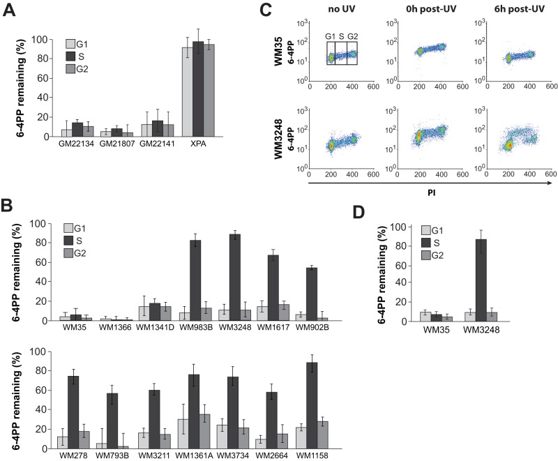
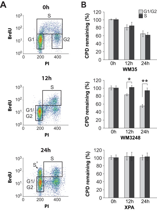
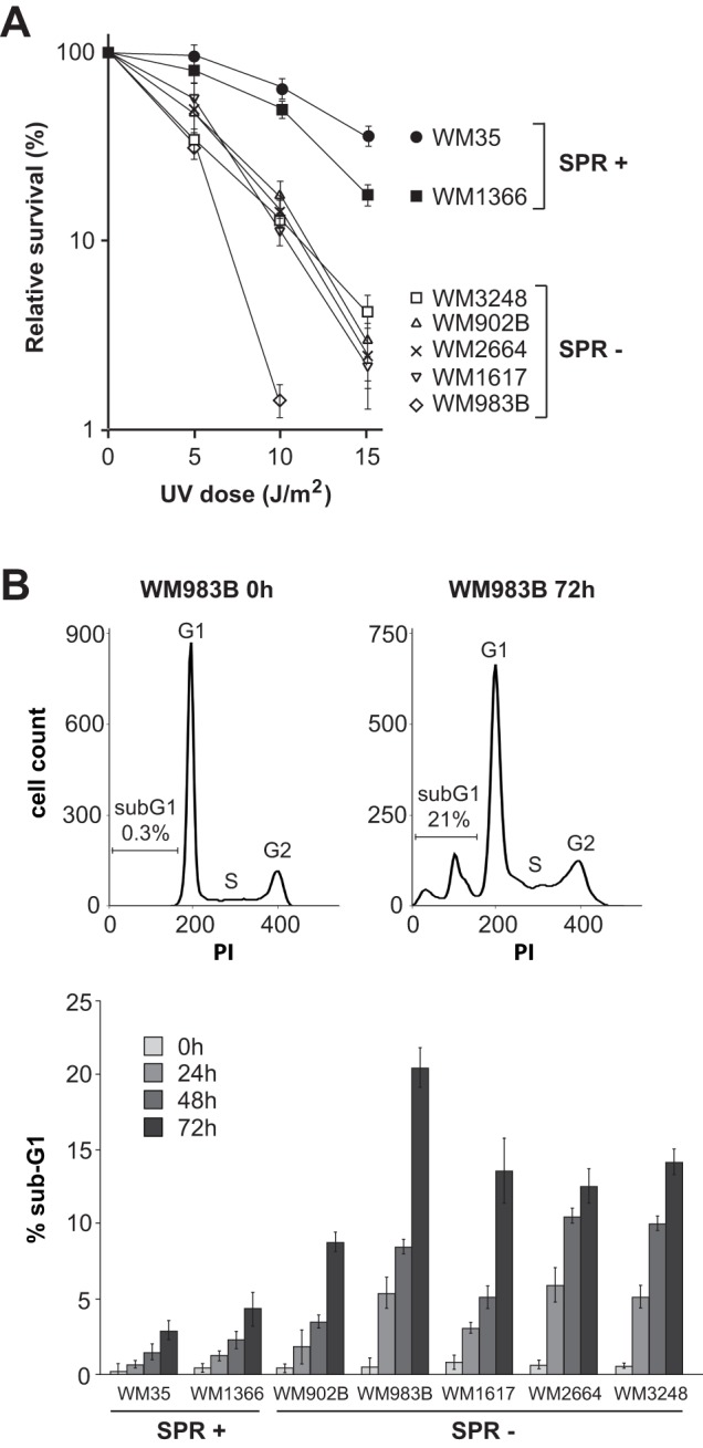
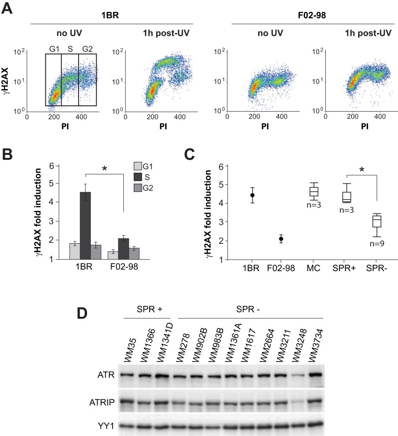
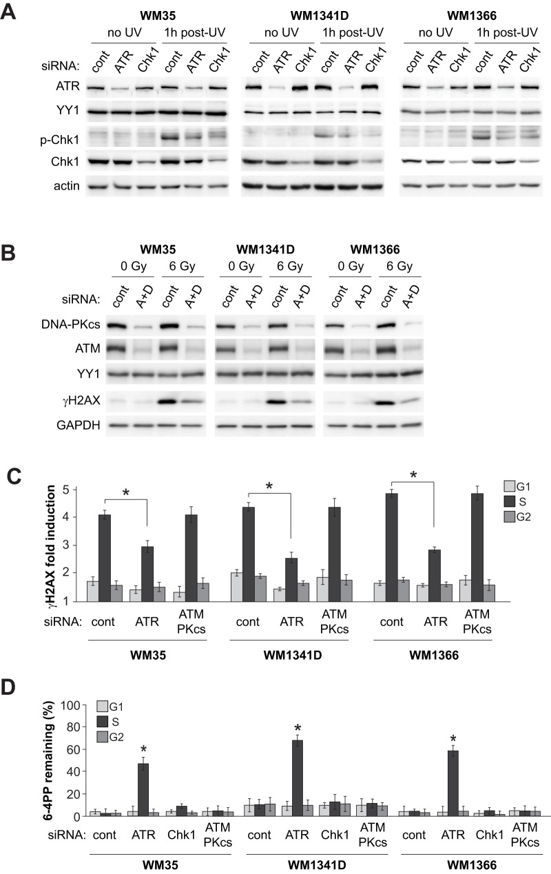
It is well established that efficient removal of highly-promutagenic UV-induced dipyrimidine photoproducts via nucleotide excision repair (NER) is required for protection against sunlight-associated malignant melanoma. Nonetheless, the extent to which reduced NER capacity might contribute to individual melanoma susceptibility in the general population remains unclear. Here we show that among a panel of 14 human melanoma strains, 11 exhibit significant inhibition of DNA photoproduct removal during S phase relative to G0/G1 or G2/M. Evidence is presented that this cell cycle-specific NER defect correlates with enhanced apoptosis and reduced clonogenic survival following UV irradiation. In addition, melanoma strains deficient in S phase-specific DNA photoproduct removal manifest significantly lower levels of phosphorylated histone H2AX at 1 h post-UV, suggesting diminished activation of ataxia telangiectasia and Rad 3-related (ATR) kinase, i.e., a primary orchestrator of the cellular response to UV-induced DNA replication stress. Consistently, in the case of DNA photoproduct excision-proficient melanoma cells, siRNA-mediated depletion of ATR (but not of its immediate downstream effector kinase Chk1) engenders deficient NER specifically during S. On the other hand simultaneous siRNA-mediated depletion of ataxia telangiectasia mutated kinase (ATM) and DNA-dependent protein kinase catalytic subunit (DNA-PKcs) exerts no significant effect on either phosphorylation of H2AX at 1 h post-UV or the efficiency of DNA photoproduct removal. Our data suggest that defective NER exclusively during S phase, possibly associated with decreased ATR signaling, may constitute an heretofore unrecognized determinant in melanoma pathogenesis.
Conflict of interest statement
Figures
Similar articles
-
DNA-PKcs plays a dominant role in the regulation of H2AX phosphorylation in response to DNA damage and cell cycle progression.BMC Mol Biol. 2010 Mar 6;11:18. doi: 10.1186/1471-2199-11-18. BMC Mol Biol. 2010. PMID: 20205745 Free PMC article.
-
ATR- and ATM-Mediated DNA Damage Response Is Dependent on Excision Repair Assembly during G1 but Not in S Phase of Cell Cycle.PLoS One. 2016 Jul 21;11(7):e0159344. doi: 10.1371/journal.pone.0159344. eCollection 2016. PLoS One. 2016. PMID: 27442013 Free PMC article.
-
Nucleotide excision repair-dependent DNA double-strand break formation and ATM signaling activation in mammalian quiescent cells.J Biol Chem. 2014 Oct 10;289(41):28730-7. doi: 10.1074/jbc.M114.589747. Epub 2014 Aug 27. J Biol Chem. 2014. PMID: 25164823 Free PMC article.
-
Xeroderma Pigmentosa Group A (XPA), Nucleotide Excision Repair and Regulation by ATR in Response to Ultraviolet Irradiation.Adv Exp Med Biol. 2017;996:41-54. doi: 10.1007/978-3-319-56017-5_4. Adv Exp Med Biol. 2017. PMID: 29124689 Free PMC article. Review.
-
Ultraviolet damage, DNA repair and vitamin D in nonmelanoma skin cancer and in malignant melanoma: an update.Adv Exp Med Biol. 2014;810:208-33. doi: 10.1007/978-1-4939-0437-2_12. Adv Exp Med Biol. 2014. PMID: 25207368 Review.
Cited by
-
Roles of UVA radiation and DNA damage responses in melanoma pathogenesis.Environ Mol Mutagen. 2018 Jun;59(5):438-460. doi: 10.1002/em.22176. Epub 2018 Feb 21. Environ Mol Mutagen. 2018. PMID: 29466611 Free PMC article. Review.
-
Know your limits: RPA availability and chemoresistance in ovarian cancer.Oncotarget. 2019 Jan 25;10(8):800-802. doi: 10.18632/oncotarget.26607. eCollection 2019 Jan 25. Oncotarget. 2019. PMID: 30783508 Free PMC article. No abstract available.
-
Targeting Rad51 as a strategy for the treatment of melanoma cells resistant to MAPK pathway inhibition.Cell Death Dis. 2020 Jul 2;11(7):581. doi: 10.1038/s41419-020-2702-y. Cell Death Dis. 2020. PMID: 32719412 Free PMC article.
-
Sun Exposure and Melanoma, Certainties and Weaknesses of the Present Knowledge.Front Med (Lausanne). 2018 Aug 30;5:235. doi: 10.3389/fmed.2018.00235. eCollection 2018. Front Med (Lausanne). 2018. PMID: 30214901 Free PMC article.
-
Hyperthermia Enhances Doxorubicin Therapeutic Efficacy against A375 and MNT-1 Melanoma Cells.Int J Mol Sci. 2021 Dec 21;23(1):35. doi: 10.3390/ijms23010035. Int J Mol Sci. 2021. PMID: 35008457 Free PMC article.
References
-
- Leiter U, Garbe C (2008) Epidemiology of melanoma and nonmelanoma skin cancer – the role of sunlight. Adv Exp Med Biol 624: 89–103. - PubMed
-
- Thompson JF, Scolyer RA, Kefford RF (2005) Cutaneous melanoma. Lancet 365: 687–701. - PubMed
-
- Gilchrest BA, Eller MS, Geller AC, Yaar M (1999) The pathogenesis of melanoma induced by ultraviolet radiation. N Engl J Med 340: 1341–1348. - PubMed
-
- Friedberg EC, Walker GC, Siede W, Wood RD, Schultz RA, et al. (Eds.) (2006) DNA Repair and Mutagenesis 2nd ed., ASM Press, Washington, DC.
Publication types
MeSH terms
Substances
LinkOut - more resources
Full Text Sources
Other Literature Sources
Research Materials
Miscellaneous
