. 2014 Jan 8;9(1):e85793.
doi: 10.1371/journal.pone.0085793. eCollection 2014.

TL1A induces TCR independent IL-6 and TNF-α production and growth of PLZF⁺ leukocytes

Affiliations

TL1A induces TCR independent IL-6 and TNF-α production and growth of PLZF⁺ leukocytes

Kirsten Reichwald et al. PLoS One. .

Abstract

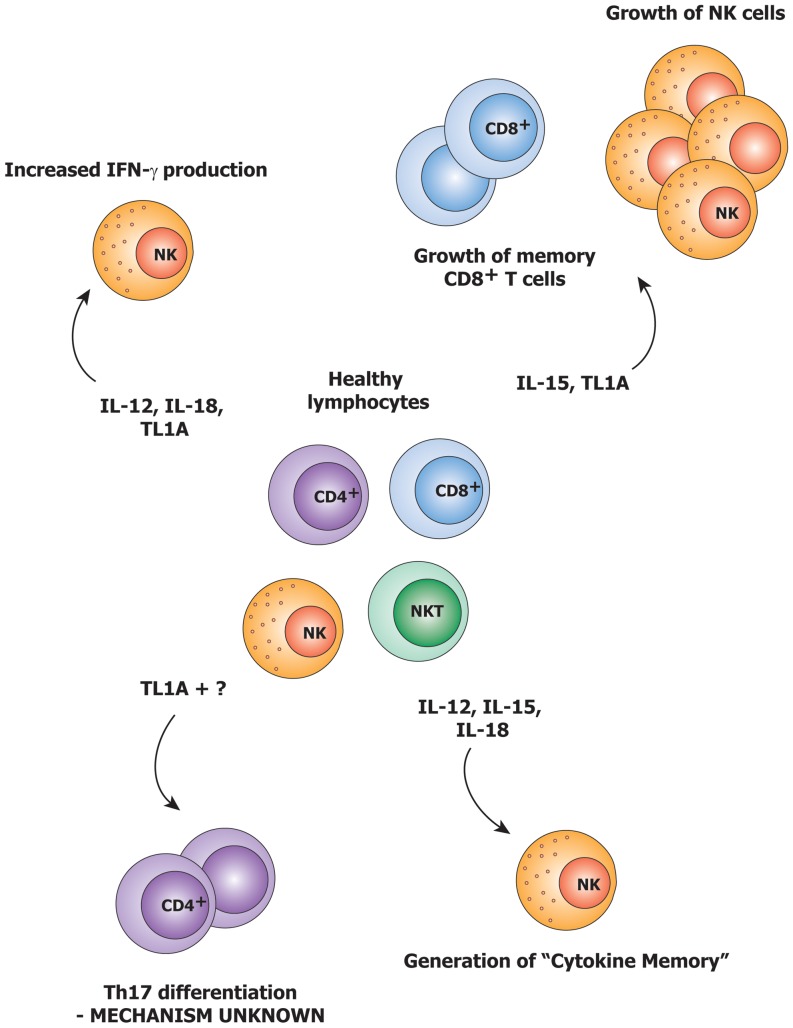
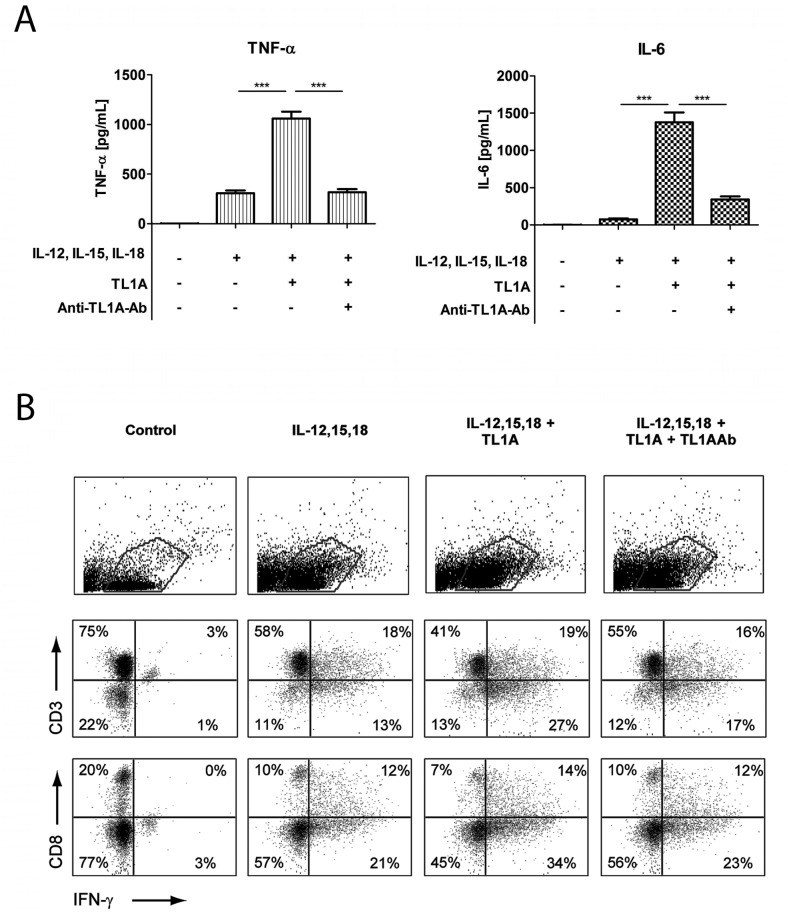
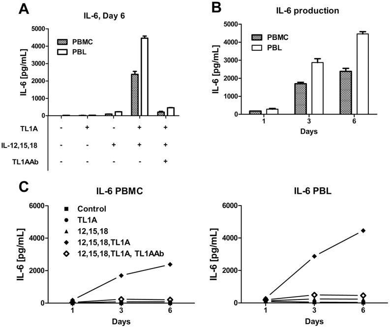
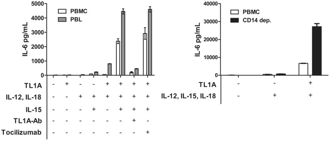
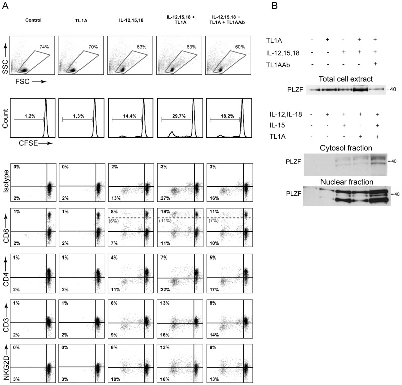
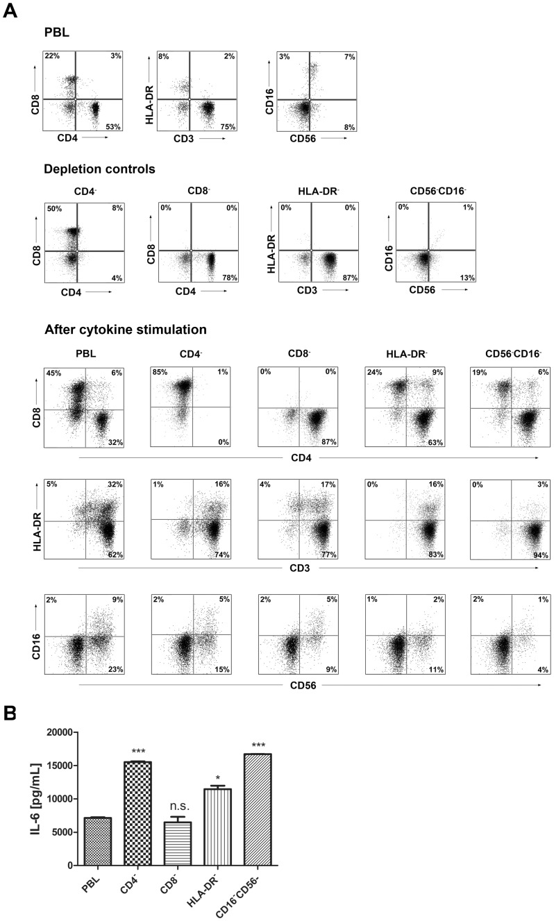
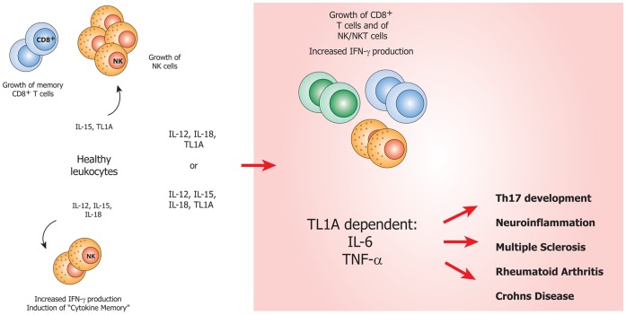
An elevated level of the cytokine TL1A is known to be associated with several autoimmune diseases, e.g. rheumatoid arthritis and inflammatory bowel disease. However, the mode of action of TL1A remains elusive. In this study, we investigated the role of TL1A in a pro-inflammatory setting, using human leukocytes purified from healthy donors. We show that TL1A, together with IL-12, IL-15 and IL-18, directly induces the production of IL-6 and TNF-α from leukocytes. Interestingly, TL1A-induced IL-6 was not produced by CD14⁺ monocytes. We further show that the produced IL-6 is fully functional, as measured by its ability to signal through the IL-6 receptor, and that the induction of IL-6 is independent of TCR stimulation. Furthermore, the transcription factor PLZF was induced in stimulated cells. These results offer a substantial explanation for the role of TL1A, since TNF-α and IL-6 are directly responsible for much of the inflammatory state in many autoimmune diseases. Our study suggests that TL1A is a possible target for the treatment of autoimmune diseases.


Conflict of interest statement

Competing Interests: The authors have declared that no competing interests exist.

Figures

Figure 1
Figure 1. An overview of previous studies on the pro-inflammatory cytokines used in our setup.
IL-15 is known to induce the growth of NK cells and memory CD8 T cells, and also causes CD8 T cells to acquire functional NK receptors , , . IL-12 and IL-18 are known to induce IFN-γ in both NK and NKT cells in synergy with TL1A . IL-12, IL-15 and IL-18 have recently been shown to induce “cytokine memory” . TL1A is also known to be involved in Th17 development , , but the mechanism remains unidentified.
Figure 2
Figure 2. TL1A induces IL-6 and TNF-α.
Freshly purified PBMCs were incubated with IL-12 (2 ng/mL), IL-15 (10 ng/mL), IL-18 (10 ng/mL), TL1A (100 ng/mL) and TL1AAb (1 µg/mL, blocking antibody). Extra IL-15 (2 ng/mL) was added on day 3. (A) After 6 days, supernatants were collected and different cytokines were measured by bead-based ELISA. Error bars represent the SEM of eight measurements. Statistically significant differences are indicated by ***(t-test, P<0.001). Data are representative of three different experiments with cells from three separate donors (B). After 6 days, cells were stained extracellularly for CD3 or CD8 and intracellularly for IFN-γ and analyzed by flow cytometry as described in the Materials and Methods. The upper panels show gating for lymphocytes; the two lower panels show CD3/IFN-γ and CD8/IFN-γ staining. Data are representative of three different experiments with cells from three separate donors.
Figure 3
Figure 3. IL-6 production is higher in PBLs than in PBMCs.
PBMCs and PBLs were prepared as described in the Materials and Methods, stimulated for 1, 3 or 6-12 (4 ng/mL), IL-15 (10 ng/mL), IL-18 (40 ng/mL), TL1A (100 ng/mL) and TL1AAb (1 µg/mL) and IL-6 was measured in the supernatants. (A) PBMCs and PBLs were stimulated with different combinations of cytokines and IL-6 was measured on day 6. (B) IL-6 production by PBMCs and PBLs stimulated with IL-12, IL-15, IL-18 and TL1A for 1, 3 and 6 days. (C) IL-6 production after 1, 3 and 6 days by PBLs and PBMCs stimulated with different cytokine combinations. Error bars represent the SEM of two measurements. Data are representative of three different experiments with cells from three separate donors.
Figure 4
Figure 4. Depletion of CD14+ monocytes enhances TL1A-dependent IL-6.
PBMCs, PBLs and CD14-depleted PBMCs were prepared as described in the Materials and Methods, stimulated for 6 days with combinations of IL-12 (4 ng/mL), IL-15 (10 ng/mL), IL-18 (40 ng/mL), TL1A (100 ng/mL), TL1AAb (1 µg/mL) and tocilizumab (RoActemra, 8 µg/mL), and IL-6 was measured in the supernatants. Error bars represent the SEM of two measurements. Data are representative of three different experiments with cells from three separate donors.
Figure 5
Figure 5. TL1A-induced IL-6 phosphorylates STAT3. A functional pSTAT3 was set up to verify the functionality of TL1A-induced IL-6.
In short, purified PBMCs were incubated for 30-6 (1, 10 or 100 ng/mL) or the supernatant from cells incubated with IL-12 (4 ng/mL), IL-15 (10 ng/mL), IL-18 (40 ng/mL), TL1A (100 ng/mL); the supernatant contained 27 ng IL-6/mL (Sup [27]). PBMC proteins were extracted and pSTAT3 and STAT3 were detected by western blotting as described. Additionally, to verify that IL-6 was in fact the cytokine responsible for STAT3 phosphorylation, both the supernatant (27 ng/mL) and rhIL-6 (10 ng/mL) were added along with tocilizumab (IL-6R blocking Ab). Data are representative of three different experiments using cells from three separate donors and supernatants from two different trials.
Figure 6
Figure 6. IL-6 production is TCR independent.
PBLs were TCR stimulated using anti-CD3 (4 µg/ml), CD3-28 beads or SEA (Staphylococcal enterotoxin A, 2.5 µg/ml). The supernatants were harvested on day 6, and bead-based ELISA was performed on the samples. Error bars represent the SEM of two measurements. Data are representative of three different experiments using PBLs from three different donors.
Figure 7
Figure 7. Intracellular IL-6 staining is possible in monocytes.
Freshly purified PBMCs were incubated for 6+/− LPS (100 ng/mL) and Golgistop or Golgiplug. Cells were stained intracellularly for IL-6, and analyzed by flow cytometry. Upper panels: monocyte gating for control and LPS samples with Golgiplug/stop. Lower panels: % of gated cells positive for IL-6 expression. Data are representative of three different experiments using PBMCs from three separate donors.
Figure 8
Figure 8. TL1A induces the growth of PLZF+ cells.
(A) PBLs were purified from healthy donors, CFSE stained and stimulated for 6 days with combinations of IL-12 (4 ng/mL), IL-15 (10 ng/mL), IL-18 (40 ng/mL), TL1A (100 ng/mL) and TL1AAb (1 µg/mL). Upper panel shows gating on live cells and (%) proliferating cells as seen by less CFSE/cell. Lower panels show staining of different surface molecules. Note that for CD8, both CD8+ (bright+dim) and CD8dim cells are shown (intermediate (%) in grey); see text for elaboration. Data are representative of three different experiments with cells from three separate donors. (B) PBLs were extracted after 6 days of stimulation with the cytokines listed, and PLZF was detected in whole cell extracts or cytoplasmic/nuclear extracts. Data are essentially representative of three different experiments.
Figure 9
Figure 9. IL-6 produced by PBLs depleted for CD4+, CD8+, HLA-DR+ or CD56+/CD16+ cells.
(A) Freshly purified PBLs stained for CD4, CD8, CD3, HLA-DR, CD16, CD56. Depletion controls for PBLs depleted for CD4+, CD8+, HLA-DR+ or CD16+/CD56+ cells. PBLs and depletions were stimulated for 7 days with combinations of IL-12 (4 ng/mL), IL-15 (10 ng/mL), IL-18 (40 ng/mL) and TL1A (100 ng/mL) and stained for CD4, CD8, CD3, HLA-DR, CD16 and CD56 expression. (B) IL-6 production after 7 days by PBLs and depletions stimulated with IL-12, IL-15, IL-18 and TL1A as described above. Error bars represent the SEM of two measurements. Statistically significant differences by t-test: *** = p<0.001, * = p<0.05, n.s. = not significant. Data are representative of two different experiments with cells from two separate donors.
Figure 10
Figure 10. Proposed role of TL1A in the induction of inflammation and Th17 pathogenesis.
IL-15 is known to support the growth of CD8+ T cells and NK cells , . The combination of IL-12, IL-15 and IL-18 induces memory-like NK-cells . We show that TL1A, in combination with IL-12 and IL-18 or IL-12, IL-15 and IL-18, specifically induces TCR-independent TNF-α and IL-6. Both are pro-inflammatory cytokines known to be key players in the development and progression of several autoimmune diseases.

Similar articles

Cited by

References

    1. Migone TS, Zhang J, Luo X, Zhuang L, Chen C, et al... (2002) TL1A is a TNF-like ligand for DR3 and TR6/DcR3 and functions as a T cell costimulator. Immunity 16: 479–492. S1074761302002832 [pii]. - PubMed
    1. Bamias G, Siakavellas SI, Stamatelopoulos KS, Chryssochoou E, Papamichael C, et al. (2008) Circulating levels of TNF-like cytokine 1A (TL1A) and its decoy receptor 3 (DcR3) in rheumatoid arthritis. Clin Immunol 129: 249–255 S1521-6616(08)00728-6 [pii];10.1016/j.clim.2008.07.014 [doi] - DOI - PubMed
    1. Cassatella MA, Pereira-da-Silva G, Tinazzi I, Facchetti F, Scapini P, et al.. (2007) Soluble TNF-like cytokine (TL1A) production by immune complexes stimulated monocytes in rheumatoid arthritis. J Immunol 178: 7325–7333. 178/11/7325 [pii]. - PubMed
    1. Zhang J, Wang X, Fahmi H, Wojcik S, Fikes J, et al. (2009) Role of TL1A in the pathogenesis of rheumatoid arthritis. J Immunol 183: 5350–5357 jimmunol.0802645 [pii];10.4049/jimmunol.0802645 [doi] - DOI - PubMed
    1. Bamias G, Evangelou K, Vergou T, Tsimaratou K, Kaltsa G, et al. (2011) Upregulation and nuclear localization of TNF-like cytokine 1A (TL1A) and its receptors DR3 and DcR3 in psoriatic skin lesions. Exp Dermatol 20: 725–731 10.1111/j.1600-0625.2011.01304.x [doi] - DOI - PubMed

Publication types

MeSH terms

Substances