Needle assisted arthroscopic clysis of the medial collateral ligament of the knee: a simple technique to improve exposure in arthroscopic knee surgery
- PMID: 24416482
- PMCID: PMC3883079
- DOI: 10.4081/or.2013.e38
Needle assisted arthroscopic clysis of the medial collateral ligament of the knee: a simple technique to improve exposure in arthroscopic knee surgery
Abstract
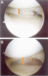
During knee arthroscopy, narrowness and tightness maybe encountered in the medial compartment that does not allow sufficient visualization or instrumentation. When this occurs, our team has found it helpful to perform a percutaneous clysis of the deep portion of the medial collateral ligament with a spinal needle. With the knee positioned in 10° to 20° of flexion and a valgus stress is applied. A spinal needle (18 Gauge) is passed percutaneously through the medial collateral ligament between the tibial plateau and undersurface of the medial meniscus. Several passes are made with the spinal needle with the bevel of the needle angled to selectively divide the fibers while keeping the medial collateral ligament under tension. Then with controlled valgus force, the medial compartment will progressively open allowing improved visualization to the posteromedial corner of the knee. This increase in space gives an enhanced visual field and further allows more room for arthroscopic instrumentation.
Keywords: knee arthroscopy; ligament release; medial collateral ligament; menisectomy.
Conflict of interest statement
Conflict of interests: the authors declare no potential conflict of interests.
Figures


Similar articles
-
The residual laxity of medial collateral ligament after magic point pie crusting MCL released in arthroscopic management of medial meniscus.Asia Pac J Sports Med Arthrosc Rehabil Technol. 2024 Sep 25;38:36-42. doi: 10.1016/j.asmart.2024.09.001. eCollection 2024 Oct. Asia Pac J Sports Med Arthrosc Rehabil Technol. 2024. PMID: 39380708 Free PMC article.
-
Arthroscopic pie-crusting release of the posteromedial complex of the knee for surgical treatment of medial meniscus injury.BMC Musculoskelet Disord. 2020 May 14;21(1):301. doi: 10.1186/s12891-020-03336-9. BMC Musculoskelet Disord. 2020. PMID: 32410610 Free PMC article.
-
[Application of pie-crusting technique of deep medial collateral ligament under arthroscopy in repairing posterior horn of medial meniscus tears].Zhongguo Gu Shang. 2021 Sep 25;34(9):840-6. doi: 10.12200/j.issn.1003-0034.2021.09.010. Zhongguo Gu Shang. 2021. PMID: 34569209 Chinese.
-
Medial collateral ligament release during knee arthroscopy: key concepts.EFORT Open Rev. 2021 Aug 10;6(8):669-675. doi: 10.1302/2058-5241.6.200128. eCollection 2021 Aug. EFORT Open Rev. 2021. PMID: 34532074 Free PMC article. Review.
-
The management of injuries to the medial side of the knee.J Orthop Sports Phys Ther. 2012 Mar;42(3):221-33. doi: 10.2519/jospt.2012.3624. Epub 2012 Feb 29. J Orthop Sports Phys Ther. 2012. PMID: 22382986 Review.
Cited by
-
The Outside-In, Percutaneous Release of the Medial Collateral Ligament for Knee Arthroscopy.Arthrosc Tech. 2020 Feb 25;9(3):e393-e397. doi: 10.1016/j.eats.2019.11.008. eCollection 2020 Mar. Arthrosc Tech. 2020. PMID: 32226748 Free PMC article.
-
The residual laxity of medial collateral ligament after magic point pie crusting MCL released in arthroscopic management of medial meniscus.Asia Pac J Sports Med Arthrosc Rehabil Technol. 2024 Sep 25;38:36-42. doi: 10.1016/j.asmart.2024.09.001. eCollection 2024 Oct. Asia Pac J Sports Med Arthrosc Rehabil Technol. 2024. PMID: 39380708 Free PMC article.
-
Which Fibers of the Medial Collateral Ligament (MCL) Should Be Released in the Pie Crust Technique Applied During Knee Arthroscopy: Superficial MCL or Deep MCL?Cureus. 2021 Dec 22;13(12):e20597. doi: 10.7759/cureus.20597. eCollection 2021 Dec. Cureus. 2021. PMID: 35103173 Free PMC article.
-
Arthroscopic release of the deep medial collateral ligament to assist in exposure of the medial tibiofemoral compartment.Arthrosc Tech. 2014 Dec 1;3(6):e699-701. doi: 10.1016/j.eats.2014.09.002. eCollection 2014 Dec. Arthrosc Tech. 2014. PMID: 25685677 Free PMC article.
-
Outside-In Deep Medial Collateral Ligament Release During Arthroscopic Medial Meniscus Surgery.Arthrosc Tech. 2016 Jul 25;5(4):e781-e785. doi: 10.1016/j.eats.2016.03.004. eCollection 2016 Aug. Arthrosc Tech. 2016. PMID: 27709037 Free PMC article.
References
-
- Jackson RW.History of arthroscopy. McGinty JB, Caspari RB, Jackson RW, Poehling GG, Operative arthroscopy. New York: Raven Press; 1991. 1-4
-
- Morin WD, Steadman JR.Arthroscopic assessment of the posterior compartments of the knee via the intercondylar notch: the arthroscopist’s field of view. Arthroscopy 1993;9:284-90 - PubMed
-
- Thijn CJ.Accuracy of double-contrast arthrography and arthroscopy of the knee joint. Skeletal Radiol 1982;8:187-92 - PubMed
-
- Warren LF, Marshall JL.The supporting structures and layers on the medial side of the knee: an anatomical analysis. J Bone Joint Surg Am 1979;61:56-62 - PubMed
-
- Gillies H, Seligson D.Precision in the diagnosis of meniscal lesions: a comparison of clinical evaluation, arthrography, and arthroscopy. J Bone Joint Surg Am 1979;61:343-6 - PubMed
LinkOut - more resources
Full Text Sources
Other Literature Sources
