

Anatomy of the human subthalamic nucleus: a combined morphometric study
- PMID: 24416591
- PMCID: PMC3876692
- DOI: 10.1155/2013/319710
Anatomy of the human subthalamic nucleus: a combined morphometric study
Abstract
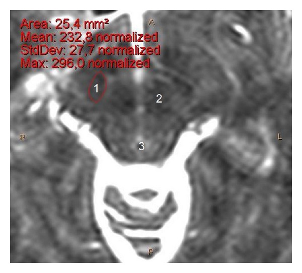
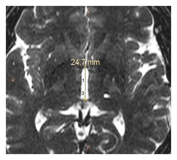
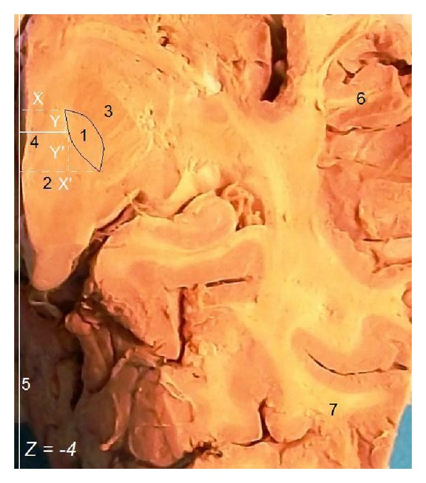
Purpose. Our purpose was to provide a combined clinically oriented study focused on the detailed anatomy of the human STN, with great respect to its targeting. Methods. For our imaging study, we used cerebral magnetic resonance images (MRIs) from 26 neurosurgical patients and for our anatomic study 32 cerebral hemispheres from 18 normal brains from cadaver donors. We measured and analyzed the STN dimensions (based on its stereotactic coordinates). Results. At stereotactic level Z = -4, the STN length was 7.7 mm on MRIs and 8.1 mm in anatomic specimens. Its width was 6 mm on MRIs and 6.3 mm in anatomic specimens. The STN was averagely visible in 3.2 transverse MRI slices and its maximum dimension was 8.5 mm. The intercommissural distance was 26.3 mm on MRIs and 27.3 mm in anatomic specimens. We found statistically significant difference of the STN width and length between individuals <60 and ≥60 years old. Conclusion. The identification of the STN limits was easier in anatomic specimens than on MRIs and easier on T2 compared to T1-weighted MRIs sections. STN dimensions appear slightly smaller on MRIs. Younger people have wider and longer STN.
Figures
Similar articles
-
Stereotactic anatomy of the human subthalamic nucleus: providing coordinates for accurate electrode placement.J Neurol Surg A Cent Eur Neurosurg. 2014 Jul;75(4):289-98. doi: 10.1055/s-0034-1368093. Epub 2014 Feb 25. J Neurol Surg A Cent Eur Neurosurg. 2014. PMID: 24570308
-
Stereotactic anatomy of the human nucleus accumbens: from applied mathematics to microsurgical accuracy.Surg Radiol Anat. 2011 Sep;33(7):583-94. doi: 10.1007/s00276-011-0804-z. Epub 2011 Mar 25. Surg Radiol Anat. 2011. PMID: 21437651
-
Anatomy of the human nucleus accumbens: a combined morphometric study.Surg Radiol Anat. 2011 Jul;33(5):405-14. doi: 10.1007/s00276-010-0766-6. Epub 2011 Jan 4. Surg Radiol Anat. 2011. PMID: 21203764
-
3T MRI evaluation of the accuracy of atlas-based subthalamic nucleus identification.Med Phys. 2008 Jul;35(7):3069-77. doi: 10.1118/1.2936229. Med Phys. 2008. PMID: 18697530
-
Magnetic resonance imaging of the subthalamic nucleus for deep brain stimulation.J Neurosurg. 2016 Jan;124(1):96-105. doi: 10.3171/2015.1.JNS142066. Epub 2015 Aug 21. J Neurosurg. 2016. PMID: 26295914 Review.
Cited by
-
Measuring Subthalamic Nucleus Volume of Parkinson's Patients and Evaluating Its Relationship with Clinical Scales at Pre- and Postdeep Brain Stimulation Treatment: A Magnetic Resonance Imaging Study.Biomed Res Int. 2021 Feb 25;2021:6646416. doi: 10.1155/2021/6646416. eCollection 2021. Biomed Res Int. 2021. Retraction in: Biomed Res Int. 2024 Mar 20;2024:9802021. doi: 10.1155/2024/9802021. PMID: 33708991 Free PMC article. Retracted. Clinical Trial.
-
The Parkinsonian Subthalamic Network: Measures of Power, Linear, and Non-linear Synchronization and their Relationship to L-DOPA Treatment and OFF State Motor Severity.Front Hum Neurosci. 2016 Oct 25;10:517. doi: 10.3389/fnhum.2016.00517. eCollection 2016. Front Hum Neurosci. 2016. PMID: 27826233 Free PMC article.
-
Patient-specific anatomical model for deep brain stimulation based on 7 Tesla MRI.PLoS One. 2018 Aug 22;13(8):e0201469. doi: 10.1371/journal.pone.0201469. eCollection 2018. PLoS One. 2018. PMID: 30133472 Free PMC article.
-
Localization of beta and high-frequency oscillations within the subthalamic nucleus region.Neuroimage Clin. 2017 Jul 24;16:175-183. doi: 10.1016/j.nicl.2017.07.018. eCollection 2017. Neuroimage Clin. 2017. PMID: 28794978 Free PMC article.
-
Psychiatric disorders after deep brain stimulation of the subthalamic nucleus in Parkinson's disease: a systematic review.Einstein (Sao Paulo). 2024 Jul 19;22:eRW0182. doi: 10.31744/einstein_journal/2024RW0182. eCollection 2024. Einstein (Sao Paulo). 2024. PMID: 39046070 Free PMC article.
References
-
- Haegelen C, Rouaud T, Darnault P, Morandi X. The subthalamic nucleus is a key-structure of limbic basal ganglia functions. Medical Hypotheses. 2009;72(4):421–426. - PubMed
-
- Acar F, Miller JP, Berk MC, Anderson G, Burchiel KJ. Safety of anterior commissure-posterior commissure-based target calculation of the subthalamic nucleus in functional stereotactic procedures. Stereotactic and Functional Neurosurgery. 2007;85(6):287–291. - PubMed
-
- Ashkan K, Blomstedt P, Zrinzo L, et al. Variability of the subthalamic nucleus: the case for direct MRI guided targeting. British Journal of Neurosurgery. 2007;21(2):197–200. - PubMed
-
- Breit S, le Bas J-F, Koudsie A, et al. Pretargeting for the implantation of stimulation electrodes into the subthalamic nucleus: a comparative study of magnetic resonance imaging and ventriculography. Neurosurgery. 2006;58(supplement 1):S83–S94. - PubMed
-
- Godinho F, Thobois S, Magnin M, et al. Subthalamic nucleus stimulation in Parkinson’s disease: anatomical and electrophysiological localization of active contacts. Journal of Neurology. 2006;253(10):1347–1355. - PubMed
LinkOut - more resources
Full Text Sources
Other Literature Sources
