. 2013:2013:620120.
doi: 10.1155/2013/620120. Epub 2013 Dec 12.

A unique case of left second supernumerary and left third bifid intrathoracic ribs with block vertebrae and hypoplastic left lung

Affiliations

A unique case of left second supernumerary and left third bifid intrathoracic ribs with block vertebrae and hypoplastic left lung

Parag Suresh Mahajan et al. Case Rep Radiol. 2013.

Abstract

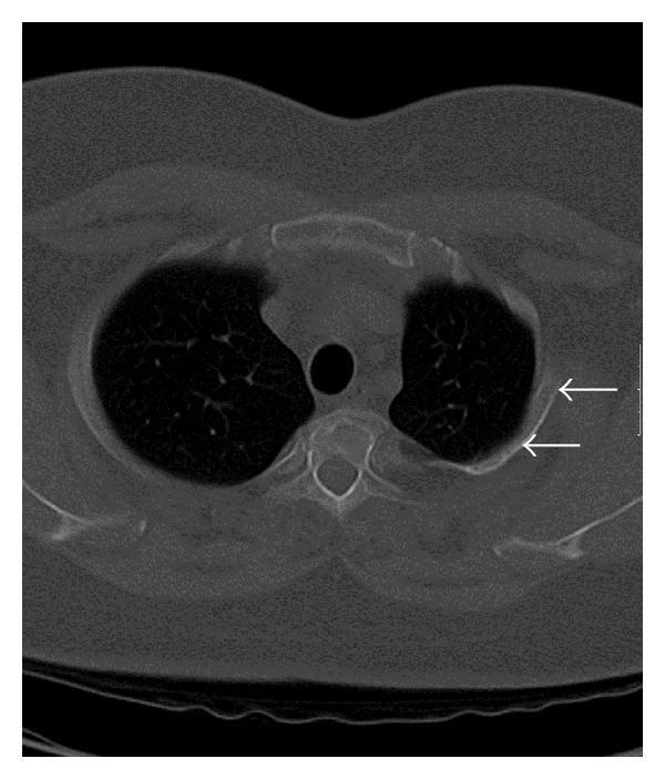
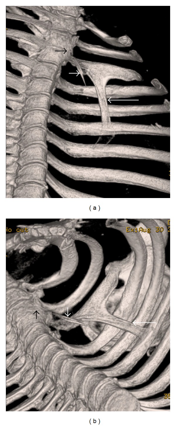
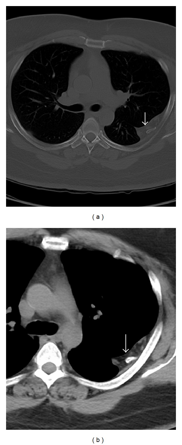
Intrathoracic rib (IR) is a very rare anomaly in which a normal, an accessory, or a bifid rib lies within the chest cavity and may originate from a vertebra or a rib. It is more commonly present on the right side, and sometimes it may be associated with vertebral anomalies. Only 50 cases have been reported to date in the literature. In most cases, the IR is an isolated finding; it is incidentally detected and is asymptomatic. The IR can be easily missed on a chest radiograph and can be mistaken initially for a pleural lesion, lung consolidation, other peripheral lung parenchymal lesions, or a bony lesion. It is, therefore, essential for physicians and radiologists to know about this entity and consider it in the differential diagnosis, to avoid further evaluation and unnecessary investigations. We present a unique case of three intrathoracic ribs, a left second supernumerary rib, left third depressed normonumerary rib, and bifid arm of the left third rib, with block vertebrae and hypoplastic left lung. To our knowledge, this is the first such case presentation in the published literature.


Figures

Figure 1
Figure 1
43-year-old gentleman complaining of high grade fever, cough, and chest pain was investigated with chest radiographs and helical chest CT scan and subsequently diagnosed with intrathoracic ribs. Chest radiograph shows left upper and mid-zone opacity (black arrows) and depression of bifid left third rib (white arrows). Upper lobe of the left lung is hypoplastic.
Figure 2
Figure 2
43-year-old gentleman complaining of high grade fever, cough, and chest pain was investigated with chest radiographs and helical chest CT scan and subsequently diagnosed with intrathoracic ribs. Unenhanced axial chest CT scan (bone window) shows depressed left third intrathoracic rib (arrows) and hypoplastic upper lobe of the left lung.
Figure 3
Figure 3
43-year-old gentleman complaining of high grade fever, cough, and chest pain was investigated with chest radiographs and helical chest CT scan and subsequently diagnosed with intrathoracic ribs. Unenhanced chest CT, volume rendered-3D images, coronal view in (a) and coronal oblique view in (b) reveal fusion of anterior aspects of the T2 and T3 vertebral bodies (short black arrow), a supernumerary second rib (thin, short, and obliquely oriented) with predominantly fibrous (non-ossified) proximal and distal parts originating from the second costovertebral neoarticulation (short white arrow), and the obliquely oriented bifid arm of the third rib coursing into the thoracic cavity (long white arrow).
Figure 4
Figure 4
43-year-old gentleman complaining of high grade fever, cough, and chest pain was investigated with chest radiographs and helical chest CT scan and subsequently diagnosed with intrathoracic ribs. Unenhanced chest CT (coronal MIP reformatting, bone window) image in (a) shows left second supernumerary (white arrow) and left third bifid (black arrow) intrathoracic ribs, prominent extrapleural soft tissue around these ribs, and hypoplastic left lung. Unenhanced chest CT (sagittal MIP reformatting, bone window) image in (b) shows left third bifid intrathoracic rib (black arrow) and prominent extrapleural soft tissue around it (white arrow).
Figure 5
Figure 5
43-year-old gentleman complaining of high grade fever, cough, and chest pain was investigated with chest radiographs and helical chest CT scan and subsequently diagnosed with intrathoracic ribs. Unenhanced axial chest CT scans with bone window in (a) and mediastinal window in (b) show prominent extrapleural soft tissue including fat around the left intrathoracic rib (arrow).

References

    1. Bottosso N, Ghaye B. Bifid intrathoracic rib. Journal Belge de Radiologie. 2008;91(3):86–87. - PubMed
    1. Kayiran SM, Gumus T, Kayiran PG, Gurakan B. Supernumerary intrathoracic rib. Archives of Disease in Childhood. 2013;98(6, article 441) - PubMed
    1. Basarslan F, Bayarogulları H, Tutanc M, Arica V, Yilmaz C, Davran R. Intrathoracic rib associated with pulmonary collapse in a pediatric patient. Iranian Journal of Radiology. 2012;9(4):220–222. - PMC - PubMed
    1. Chung JH, Pipavath SN. Images in clinical medicine. Intrathoracic rib. New England Journal of Medicine. 2009;361(26, article 2557) - PubMed
    1. Apaydin M, Sarsilmaz A, Varer M. Third accessory (supernumerary) intrathoracic right rib. Surgical and Radiologic Anatomy. 2009;31(8):641–643. - PubMed

LinkOut - more resources