

PD-L1 expression in the Merkel cell carcinoma microenvironment: association with inflammation, Merkel cell polyomavirus and overall survival
- PMID: 24416729
- PMCID: PMC3885978
- DOI: 10.1158/2326-6066.CIR-13-0034
PD-L1 expression in the Merkel cell carcinoma microenvironment: association with inflammation, Merkel cell polyomavirus and overall survival
Abstract
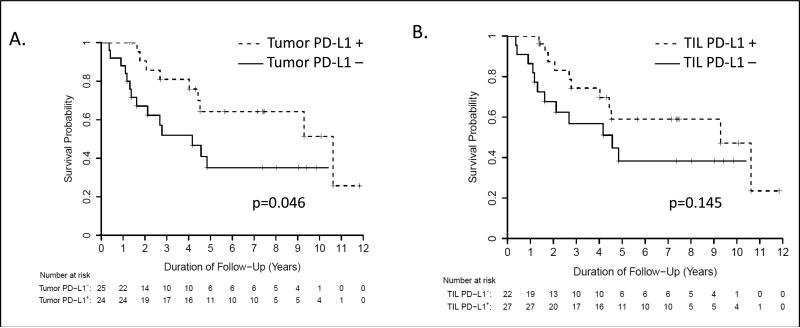
Merkel cell carcinoma (MCC) is a lethal, virus-associated cancer that lacks effective therapies for advanced disease. Agents blocking the PD-1/PD-L1 pathway have demonstrated objective, durable tumor regressions in patients with advanced solid malignancies and efficacy has been linked to PD-L1 expression in the tumor microenvironment. To investigate whether MCC might be a target for PD-1/PD-L1 blockade, we examined MCC PD-L1 expression, its association with tumor-infiltrating lymphocytes (TILs), Merkel cell polyomavirus (MCPyV), and overall survival. Sixty-seven MCC specimens from 49 patients were assessed with immunohistochemistry for PD-L1 expression by tumor cells and TILs, and immune infiltrates were characterized phenotypically. Tumor cell and TIL PD-L1 expression were observed in 49% and 55% of patients, respectively. In specimens with PD-L1(+) tumor cells, 97% (28/29) demonstrated a geographic association with immune infiltrates. Among specimens with moderate-severe TIL intensities, 100% (29/29) demonstrated PD-L1 expression by tumor cells. Significant associations were also observed between the presence of MCPyV DNA, a brisk inflammatory response, and tumor cell PD-L1 expression: MCPyV(-) tumor cells were uniformly PD-L1(-). Taken together, these findings suggest that a local tumor-specific and potentially MCPyV-specific immune response drives tumor PD-L1 expression, similar to previous observations in melanoma and head and neck squamous cell carcinomas. In multivariate analyses, PD-L1(-) MCCs were independently associated with worse overall survival (hazard ratio 3.12; 95% CI, 1.28-7.61; p=0.012). These findings suggest that an endogenous immune response promotes PD-L1 expression in the MCC microenvironment when MCPyV is present, and provide a rationale for investigating therapies blocking PD-1/PD-L1 for patients with MCC.
Keywords: B7-H1; Merkel cell carcinoma; Merkel cell polyomavirus; PD-L1; immunotherapy.
Figures


References
Publication types
MeSH terms
Substances
Grants and funding
LinkOut - more resources
Full Text Sources
Other Literature Sources
Medical
Research Materials
