. 2014 Apr;146(4):1028-39.
doi: 10.1053/j.gastro.2014.01.015. Epub 2014 Jan 11.

Mutations in tetratricopeptide repeat domain 7A result in a severe form of very early onset inflammatory bowel disease

Affiliations

Mutations in tetratricopeptide repeat domain 7A result in a severe form of very early onset inflammatory bowel disease

Yaron Avitzur et al. Gastroenterology. 2014 Apr.

Abstract

Background & aims: Very early onset inflammatory bowel diseases (VEOIBD), including infant disorders, are a diverse group of diseases found in children younger than 6 years of age. They have been associated with several gene variants. Our aim was to identify the genes that cause VEOIBD.

Methods: We performed whole exome sequencing of DNA from 1 infant with severe enterocolitis and her parents. Candidate gene mutations were validated in 40 pediatric patients and functional studies were carried out using intestinal samples and human intestinal cell lines.

Results: We identified compound heterozygote mutations in the Tetratricopeptide repeat domain 7 (TTC7A) gene in an infant from non-consanguineous parents with severe exfoliative apoptotic enterocolitis; we also detected TTC7A mutations in 2 unrelated families, each with 2 affected siblings. TTC7A interacts with EFR3 homolog B to regulate phosphatidylinositol 4-kinase at the plasma membrane. Functional studies demonstrated that TTC7A is expressed in human enterocytes. The mutations we identified in TTC7A result in either mislocalization or reduced expression of TTC7A. Phosphatidylinositol 4-kinase was found to co-immunoprecipitate with TTC7A; the identified TTC7A mutations reduced this binding. Knockdown of TTC7A in human intestinal-like cell lines reduced their adhesion, increased apoptosis, and decreased production of phosphatidylinositol 4-phosphate.

Conclusions: In a genetic analysis, we identified loss of function mutations in TTC7A in 5 infants with VEOIBD. Functional studies demonstrated that the mutations cause defects in enterocytes and T cells that lead to severe apoptotic enterocolitis. Defects in the phosphatidylinositol 4-kinase-TTC7A-EFR3 homolog B pathway are involved in the pathogenesis of VEOIBD.

Keywords: Autoimmunity; IBD; Intestinal Atresia; Intestine.


Conflict of interest statement

Conflict of Interest: none

Figures
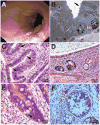
Figure 1
Figure 1. Histological and Endoscopic Characteristics of Intestine of Patients with TTC7A Mutations
Figure 1A. Colonoscopy showed severe inflammation characterized by continuous grade 2 colitis and multiple areas of exfoliation and sloughing of the surface epithelium from Family-1. Figure 1B. Low magnification electron micrograph of crypt epithelium from the same biopsy as shown in C from Family-1. Amongst regenerating crypt cells, apoptotic cell (*), enteroendocrine cell (EC) and Paneth cell (PC) is present. Crypt enterocytes showed sparse brush border microvilli (arrow) (Scale bar 10 μm). Figure 1C. High magnification of duodenal crypt epithelium showed extensive apoptosis from Family 1 (arrows) (H & E stain; Scale bar on Figure). Figure 1D–E. Low (E) and High (F) magnification of Cecum epithelium showed extensive apoptosis from Family-2 (arrows) (H & E stain). Figure 1F. High (F) magnification of Cecum epithelium showed extensive apoptosis from Family-3 (H & E stain).
Figure 2
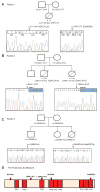
Figure 2. TTC7AGenetic Analysis
Figure 2A. Pedigree and TTC7A Mutations in Family-1. Patient from Family 1was heterozygous for 211G>A (p.E71K) inherited from her father and c.1944 C>T (p.Q526X) inherited from her mother. Figure 2B. Pedigree and TTC7A Mutations in Family-2. Siblings from Family 2 were heterozygous for a novel c.844-1 G>T TTC7A mutation in splice acceptor site of exon 7 inherited from the mother and a novel c.1204-2 A>G TTC7A mutation in splice acceptor site of exon 10 inherited from the father. A third sibling was found to be negative for both mutations. Figure 2C. Pedigree and TTC7A Mutations in Family-3. Siblings from Family 2 were homozygous for a non-synonymous mutation in exon 20 at c.G2494A resulted in a alanine acid to threonine substitution at amino acid position 832 (p.A832T). Figure 2D. Location of TTC7A Mutations in Patients. Cartoon of TTC7A protein with TPR domains in red and identified mutations highlighted.
Figure 3
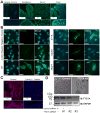
Figure 3. Functional TTC7A Enterocyte Studies
Figure 3A. TTC7A Expression in Intestinal Enterocytes. Immunofluorescence microscopy performed on human tissue sections immunostained using anti-TTC7A antibody (and DAPI) demonstrates TTC7A expression in enterocytes within the duodenum, ileum and colon. Lower inset panels represent zoomed images of the corresponding panel above. Scale bars = 100 μm. Figure 3B. E71K, Q526X and A832T Mutations in TTC7A. Caco-2 cells were transiently transfected with Myc-tagged wild-type, E71K, Q526X, and A832T TTC7A, immunostained using anti-Myc antibody and visualized using confocal microscopy. Negative control panels represent staining with secondary antibody only. The control plasmid represents an empty vector sham transfection. Scale bars = 25μm. Figure 3C. TTC7A in Enterocytes from Patient Cecum (Family 2) Immunofluorescence microscopy was performed on TTC7A-immunostained (and DAPI) cecal tissues sections from both control and patient (Family 2) biopsies. Compared to control staining (left panel), TTC7A expression is reduced in the patient sample (right panel). Scale bar = 100μm. Figure 3D. Stable knockdown of TTC7A Resulted in Morphological Changes in Henle-407 cells. Expression of TTC7A in stably transfected Henle-407 cells was reduced (70–80%) compared to Henle-407 cells stably transfected with control shRNA. The impact on cellular morphology of control and TTC7A shRNA knockdown was examined in Henle 407 cells by contrast microscopy (100X magnification) under normal culture conditions. Knockdown of TTC7A resulted in a loss of cobblestone morphology and development of fibroblastoid morphology with spindle-like features.
Figure 4
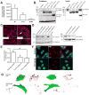
Figure 4
Figure 4A. Impaired Cell Adhesion in TTC7A-depleted Cells. The total dissociation time, defined as the time required for complete dissociation of all cells from the tissue culture plate, was markedly reduced in TTC7A-depleted Henle-407 cells compared to control cells. Dissociation Assay (N=6 biological replicates, Student’s t-test, p = 0.0022) Figure 4B. Impaired Cell Adhesion to Collagen and Fibronectin in TTC7A-depleted cells. Cell adhesion assays were performed using crystal violet staining. Control and TTC7A-depleted Henle 407 cells were seeded on 96 well plates coated with either collagen or fibronectin. Adhesion was assessed on the basis of optical density (OD) at 570nm. N=3, (*) Adhesion Assay N=3 biological replicates, Student’s t-test, p(Collagen) = 0.027 and p(Fibronectin) = 0.0077 Figure 4C. TTC7A-depletion in Henle 407 Cells Results in Greater Caspase-dependent Apoptosis. To investigate the impact of TTC7A suppression on the induction of apoptosis, the activation of caspase-3 was measured by western blot. Specific cleavage of pro-caspase 3 (32 kDa) into the active caspase-3 fragments (17k Da) was increased in cells serum-starved for 24 and 48 hours. N = 3, (*) p = 0.012, ANOVA Figure 4D. TTC7A-depletion in Henle 407 Cells Results in Greater Apoptosis Measured by Flow Cytometric Analysis of Annexin V. To examine the significance of TTC7A suppression, following loss of attachment, flow cytometric analysis was conducted to quantity the extent of apoptosis in cells starved for 24 and 48 hours. Cells were stained with AnnexinV-PE and 7-AAD (viability marker); apoptotic cells were identified as AnnexinV+ 7-AAD cells. In TTC7A-depleted cells, the proportion of cells in early apoptosis increased to approximately 4.5% at 24 hours and 11.2% at 48 hours of serum-starvation compared to 1.3% (24 hours) and 4.1% (48 hours) in control cells. Annexin V Apoptosis Assay N=3 biological replicates, Student’s t-test, p(FBS, 24h = 0.0018, p(FBS,48h) = 0.0076, p(SS,24) = 0.021, p(SS,48h) = 0.0034
Figure 5
Figure 5
Figure 5A. Tandem Mass Spectrometry. E71K and Q526X mutations reduce the ability of TTC7A to immunoprecipitate PI4KA. Selected peptides from PI4KA and TTC7A were analyzed to determine the area under their MS1 peaks to assess the relative abundance of each peptide. The PI4KA present in each sample was normalized to the total TTC7A in each technical replicate to allow comparisons among biological replicates (n=3). These normalized values were averaged over all experiments. Error bars represent the standard error. Figure 5B. PI4KIIIα-TTC7A Co-immunoprecipitate. HEK293T cells were transiently transfected with Myc-tagged wild type (WT-TTC7A), E71K, Q526X, and A832T TTC7A constructs. Lysates were immunoprecipated with anti-Myc antibody, and then immunoblotted using anti-PI4KIIIα and anti-Myc (for TTC7A) antibodies. The control lane represents transfection with an empty vector. Figure 5C. Expression and Localization of PI4KIIIα is Altered in Patients with TTC7A-Deficiency Immunofluorescence microscopy was performed on both control and patient colonic tissue sections immunostained with anti-PI4KIIIα antibodies. In the left panel, immunohistochemistry demonstrated that PI4KIIIα is highly expressed in enterocytes and immune cells from healthy human intestine. Inset panel depicts zoomed view of Panel 1 demonstrating PI4KIIIα expression at the plasma membrane of enterocytes. In the patient tissues, immunohistochemistry demonstrated that PI4KIIIα is dysregulated in enterocytes. Inset panel (representing region indicated by white arrow) demonstrates loss of PI4KIIIα at the plasma membrane of enterocytes bordering the intestinal crypt. Scale bar = 100μm. Figure 5D. shRNA-mediated knockdown of TTC7A expression leads to decreased PI4KIIIα levels To test the efficacy of the TTC7A shRNA, Henle-407 cells were transiently co-transfected with wild-type TTC7A and the various knock-down constructs, labelled #1 through #4, including a scrambled shRNA control and sham transfection. shRNA #1 and #3 showed reduction in TTC7A expression (left panels). shRNA containing the same targeting sequences were used to lentivirally infect Henle-407 cells where expression of PI4KIIIα was assessed in cell lysates by Western blot (right panels). GAPDH was stained as loading control for all blots. Figure 5E. Quantitation of PI4KIIIα expression in Henle-407 cells infected with TTC7A shRNA. Quantitation of Western blot band intensities from Figure 5D demonstrates a statistically significant reduction in PI4KIIIα expression following TTC7A knockdown (Student’s t-test, n = 3, p = 0.0234). Figure 5Fi, Fii. TTC7A Depletion Results in Decreased PtdIns-4P (PI-4P) Production. (Fi – Cytoplasmic PIP) TTC7A Knockdown and Control Henle-407 cells were stained with antibodies against Ptdlns4P (in red; Z-P004, IgM, Cedarlane, USA) and DAPI (in blue) to visualize nuclei. (Fii – Plasma Membrane PI-4P) TTC7A knockdown Henle-407 cells have reduced plasma membrane immunostaining for PI-4P compared to controls. For control and TTC7A knockdown (KD), Henle-407 cells Z-stack images were generated at 0.2μm intervals and recapitulated using Volocity to generate a three-dimensional model to illustrate cell surface levels of PIP. Unconjugated GFP, expressed from the control and knockdown plasmids, was visualized and used to approximate the morphology of the cells. Each pair of images represents two views of the same cell according to axes depicted.
Figure 6
Figure 6. Summary of TTC7 Mutations
Schematic representation of the role of TTC7A in the trafficking of PI4KIIIα to the plasma membrane from the trans-Golgi network. The left panel represents wild-type TTC7A in enterocytes wherein TTC7A binds to and facilitates the transport of PI4KIIIα from the trans-Golgi to the plasma membrane. At the membrane, PI4KIIIα can catalyze the production of PtdIns-4P(PI-4P). PI-4P levels at the plasma membrane have been implicated in cell survival and the maintenance of cell polarity. In the right panel, the various TTC7A mutations identified in the patients are depicted. E71K, Q526X and A832T TTC7A all demonstrated reduced binding to PI4KIIIα which could reduce the interaction between TTC7A and PI4KIIIα, hindering transport to the PM. Consequently, this will lead to reduced plasma membrane levels of PI-4P, a dysregulation that would affect downstream signaling pathways.

References

    1. Muise AM, Snapper SB, Kugathasan S. The age of gene discovery in very early onset inflammatory bowel disease. Gastroenterology. 2012;143:285–8. - PubMed
    1. Griffiths AM. Specificities of inflammatory bowel disease in childhood. Best Pract Res Clin Gastroenterol. 2004;18:509–23. - PubMed
    1. Heyman MB, Kirschner BS, Gold BD, et al. Children with early-onset inflammatory bowel disease (IBD): analysis of a pediatric IBD consortium registry. J Pediatr. 2005;146:35–40. - PubMed
    1. Glocker EO, Kotlarz D, Boztug K, et al. Inflammatory bowel disease and mutations affecting the interleukin-10 receptor. N Engl J Med. 2009;361:2033–45. - PMC - PubMed
    1. Kotlarz D, Beier R, Murugan D, et al. Loss of interleukin-10 signaling and infantile inflammatory bowel disease: implications for diagnosis and therapy. Gastroenterology. 2012;143:347–55. - PubMed

Publication types

MeSH terms

Supplementary concepts