. 2014 Mar 6:1550:18-26.
doi: 10.1016/j.brainres.2014.01.002. Epub 2014 Jan 10.

Corneal pain activates a trigemino-parabrachial pathway in rats

Affiliations

Corneal pain activates a trigemino-parabrachial pathway in rats

Sue A Aicher et al. Brain Res. .

Abstract

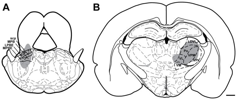
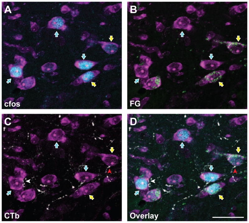
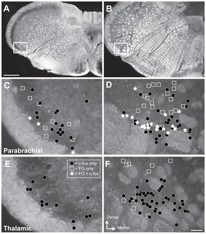
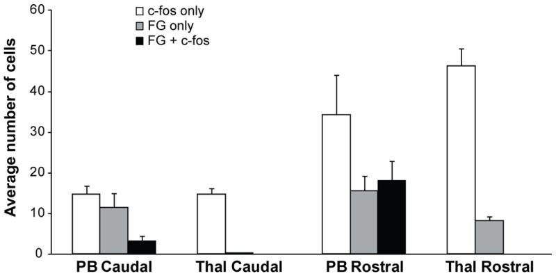
Corneal pain is mediated by primary afferent fibers projecting to the dorsal horn of the medulla, specifically the trigeminal nucleus caudalis. In contrast to reflex responses, the conscious perception of pain requires transmission of neural activity to higher brain centers. Ascending pain transmission is mediated primarily by pathways to either the thalamus or parabrachial nuclei. We previously showed that some corneal afferent fibers preferentially contact parabrachial-projecting neurons in the rostral pole of the trigeminal nucleus caudalis, but the role of these projection neurons in transmitting noxious information from the cornea has not been established. In the present study, we show that noxious stimulation of the corneal surface activates neurons in the rostral pole of the nucleus caudalis, including parabrachially projecting neurons that receive direct input from corneal afferent fibers. We used immunocytochemical detection of c-Fos protein as an index of neuronal activation after noxious ocular stimulation. Animals had previously received injections of a retrograde tracer into either thalamic or parabrachial nuclei to identify projection neurons in the trigeminal dorsal horn. Noxious stimulation of the cornea induced c-Fos in neurons sending projections to parabrachial nuclei, but not thalamic nuclei. We also confirmed that corneal afferent fibers identified with cholera toxin B preferentially target trigeminal dorsal horn neurons projecting to the parabrachial nucleus. The parabrachial region sends ascending projections to brain regions involved in emotional and homeostatic responses. Activation of the ascending parabrachial system may explain the extraordinary salience of stimulation of corneal nociceptors.

Keywords: Cholera toxin B; Cornea; Noxious; Parabrachial; Thalamus.


Figures

Figure 1
Figure 1
Schematic representation of FluoroGold injection sites into left parabrachial nuclei (A) or right thalamic nuclei (B) that yielded retrograde labeling in the left trigeminal dorsal horn (Paxinos and Watson atlas, with permission). Abbreviations: scp = superior cerebellar peduncle, MPB = medial parabrachial nucleus, LPBE = lateral parabrachial nucleus, external, MPBE = medial parabrachial nucleus, external. LDVL = laterodorsal thalamic nuclei, ventrolateral, Po = posterior thalamic nuclear group, VPM = ventral postermedial thalamic nuclei, VPL = ventrolateral posterolateral thalamic nuclei, VL = ventrolateral thalamic nuclei, VM = ventromedial thalamic nuclei. Scale bar = 1 mm
Figure 2
Figure 2
FluoroGold and c-Fos are co-localized in subsets of trigeminal dorsal horn neurons that also receive corneal afferent input. Micrographs of the same area showing the detection of individual markers are shown in panels A – C, while panel D shows all three channels in a single micrograph. A. Neurons were identified with NeuroTrace fluorescent Nissl stain (magenta). A subset of these neurons are immunoreactive for c-Fos (blue; blue arrows) following noxious stimulation of the cornea. B. Immunoreactivity for FluoroGold (green) shows a distinct subset of neurons containing both c-Fos and FluoroGold (yellow arrows). Thus, these FG-containing projection neurons are also activated by noxious corneal stimulation. C. CTb-containing corneal afferents (white) are seen in apposition to select subpopulations of trigeminal dorsal horn neurons. A neuron that receives a CTb apposition and also contains FG and c-Fos is indicated with a red arrowhead. D. Overlay of all four channels showing CTb appositions with cells containing both FG and c-Fos. Each panel is composed of a Z stack of 10 consecutive images for total depth of 3.6 μm. Scale bar = 25 μm
Figure 3
Figure 3
Projection neurons are interspersed with neurons activated by noxious ocular stimulation in trigeminal nucleus caudalis. Representative distribution of retrogradely labeled (FG) and c-Fos-immunoreactive (-ir) neurons within caudal and rostral regions of ventrolateral nucleus caudalis (Vc) that were analyzed in the present studies. A, B. Darkfield micrographs depicting the regions of interest for caudal Vc (A) and rostral Vc (B). White box shows the region examined in the analysis. C – F. Drawings of distribution of labeled neurons in representative cases. The area of interest contained neurons with FG only (open squares), c-Fos only (dark circles) or both (white stars) for injections into parabrachial (C, D) or thalamic (E, F) nuclei. Results show that c-Fos-ir neurons (black circles) are more abundant in rostral Vc (D, F) compared to caudal Vc in all cases. Injections into parabrachial nuclei produced more retrogradely labeled neurons (open squares; C, D) than injections into thalamic nuclei (E, F); and only parabrachial cases had retrogradely labeled neurons that also contained c-Fos (white stars; C, D) after noxious corneal stimulation. Scale bar = 500 μm for panels A and B, and 50 μm for panels C - F
Figure 4
Figure 4
Quantification of cell counts within caudal and rostral levels of trigeminal nucleus caudalis (Vc) for cases that received retrograde tracer (FG) injections into either the parabrachial nuclei (PB) or thalamic nuclei (Thal). Each cell count classification is mutually exclusive. Cells containing c-Fos only (white bars) were detected in all cases in both caudal and rostral Vc, but were more abundant within rostral Vc. Cells containing FG only (gray bars) were detected in the caudal Vc for PB cases, but not for Thal cases. Cells containing FG only (gray bars) were present in rostral Vc for both PB and Thal cases, indicating that some projection neurons were not activated by the noxious stimuli applied to the cornea. Projection neurons that were activated by noxious stimulation of the cornea (FG + c-Fos; black bars) were only present in PB cases and were seen in both caudal and rostral Vc. In rostral Vc, more than half of the PB-projecting neurons identified were activated by noxious stimulation of the cornea (black versus gray bars).

References

    1. Acosta MC, Belmonte C, Gallar J. Sensory experiences in humans and single-unit activity in cats evoked by polymodal stimulation of the cornea. J Physiol. 2001;534:511–525. - PMC - PubMed
    1. Aicher SA, Hermes SM, Hegarty DM. Corneal afferents differentially target thalamic- and parabrachial-projecting neurons in spinal trigeminal nucleus caudalis. Neuroscience. 2013;232:182–193. - PMC - PubMed
    1. Aicher SA, Mitchell JL, Swanson KC, Zadina JE. Endomorphin-2 axon terminals contact mu-opioid receptor-containing dendrites in trigeminal dorsal horn. Brain Res. 2003;977:190–198. - PubMed
    1. Aicher SA, Reis DJ, Nicolae R, Milner TA. Monosynaptic projections from the medullary gigantocellular reticular formation to sympathetic preganglionic neurons in the thoracic spinal cord. J Comp Neurol. 1995;363:563–580. - PubMed
    1. Allen GV, Barbrick B, Esser MJ. Trigeminal-parabrachial connections: Possible pathway for nociception-induced cardiovascular reflex responses. Brain Res. 1996;715:125–135. - PubMed

Publication types

MeSH terms

Substances