. 2014 Jan 20:449:140-9.
doi: 10.1016/j.virol.2013.10.037. Epub 2013 Dec 5.

Analysis of the N-terminal positively charged residues of the simian immunodeficiency virus Vif reveals a critical amino acid required for the antagonism of rhesus APOBEC3D, G, and H

Affiliations

Analysis of the N-terminal positively charged residues of the simian immunodeficiency virus Vif reveals a critical amino acid required for the antagonism of rhesus APOBEC3D, G, and H

Kimberly Schmitt et al. Virology. .

Abstract

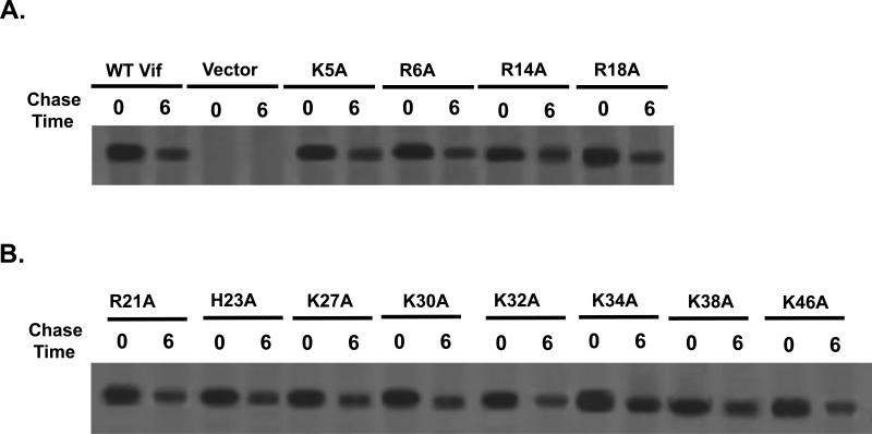
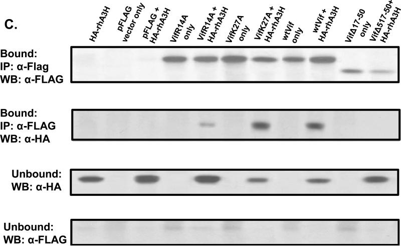
Previous studies have shown that apolipoprotein B mRNA editing, enzyme catalytic, polypeptide G (APOBEC3G; hA3G) and F (APOBEC3F; hA3F) proteins interact with a nonlinear binding site located at the N-terminal region of the HIV-1 Vif protein. We have analyzed the role of 12 positively charged amino acids of the N-terminal region of the SIV Vif. Simian-human immunodeficiency viruses (SHIV) were constructed that expressed each of these amino acid substitutions. These viruses were examined for replication in the presence of rhesus macaque APOBEC3 proteins (rhA3A-rhA3H), incorporation of the different A3 proteins into virions, and replication in rhesus macaque PBMC. Similar to other studies, we found that K27 was essential for rhA3G activity and rhA3F but was not important for restriction of SHIVΔvif by rhA3A, rhA3D or rhA3H. Our results identified the arginine at position 14 of the SIV Vif as a critical residue for virus restriction by rhA3D, rhA3G and rhA3H.

Keywords: APOBEC3 proteins; Amino terminus; HIV-1; SHIV; SIV; Structure–function; Vif; Virus restriction.


Figures

Fig. 1
Fig. 1
Alignment of the amino terminal 50 amino acids of the HIV-1 and SIV Vif proteins. Underneath the SIV Vif sequence, the mutants analyzed in this study are indicated in bold.
Fig. 2
Fig. 2
Expression of the different mutant Vif proteins. Vectors expressing each Flag-tagged Vif mutant were transfected into 293 cells for 48 h. The cells were starved for methionine/cysteine and radiolabeled with 35S-methionine/cysteine for 60 minutes. The radiolabel was removed, the cells washed three times, and incubated in medium containing cold excess methionine/cysteine for either 0 or 6 h. The cells were lysed in 1X RIPA buffer and the FLAG-tag containing Vif proteins immunoprecipitated with a rabbit anti-Flag antibody. Panel A. Results of the pulse-chase analyses of cells transfected with vectors expressing the unmodified Vif, Δvif (first 27 amino acids), VifK5A, VifR6A, VifK14A, and VifR18A. Panel B. Results of the pulse-chase analyses of cells transfected with vectors expressing the VifK21A, VifH23A, VifK27A, VifK30A, VifK32A, VifK34A, VifK38A, and K46A. The time of the chase in hours is indicated above each lane.
Fig. 3
Fig. 3
Replication of SHIVs expressing each Vif mutant in SupT1 cells. SupT1 cells were inoculated with equivalent levels (25 ng) of SHIVKU-2MC4, SHIVΔvif, or viruses expressing each of the mutant Vif proteins. At 4 h post-inoculation, the medium was removed and replaced with fresh medium and incubated for 15 days. Replication was assessed by the level of p27 in the medium at specific days post-inoculation. The experiment was performed twice and p27 levels did not vary by more than 5%. Panel A. Replication of SHIVKU-2MC4, SHIVΔvif, and SHIVs expressing Vif mutants R5A, K6A, R14A, K18A, R21A, and H23A. Panel B. Replication of SHIVKU-2MC4, SHIVΔvif, and SHIVs expressing Vif mutants K27A, K30A, K32A, K34A, K38A, and K46A.
Fig. 3
Fig. 3
Replication of SHIVs expressing each Vif mutant in SupT1 cells. SupT1 cells were inoculated with equivalent levels (25 ng) of SHIVKU-2MC4, SHIVΔvif, or viruses expressing each of the mutant Vif proteins. At 4 h post-inoculation, the medium was removed and replaced with fresh medium and incubated for 15 days. Replication was assessed by the level of p27 in the medium at specific days post-inoculation. The experiment was performed twice and p27 levels did not vary by more than 5%. Panel A. Replication of SHIVKU-2MC4, SHIVΔvif, and SHIVs expressing Vif mutants R5A, K6A, R14A, K18A, R21A, and H23A. Panel B. Replication of SHIVKU-2MC4, SHIVΔvif, and SHIVs expressing Vif mutants K27A, K30A, K32A, K34A, K38A, and K46A.
Fig. 4
Fig. 4
The incorporation of A3 proteins into viruses expressing each of the mutant Vif proteins. 293 cells were transfected with a plasmids expressing rhesus macaque tagged A3 proteins and genomes for SHIVKU-2MC4, SHIVΔvif, or the SHIVs expressing the different mutant Vif proteins. At 24 h post-transfection, the cells were starved for methionine/cysteine and radiolabeled with 35S-methionine/cysteine for 12 h. The culture medium was collected and the virus partially purified as described in the Materials and Methods section. The virus preparations were lysed in 1X RIPA buffer. Half of the samples were immunoprecipitated with an anti-HA serum to detect the HA-tag containing A3 proteins while the other half immunoprecipitated with anti-SHIV serum to detect the p27 protein. Samples were normalized for equivalent amounts of p27. A3 proteins were also immunoprecipitated from cell lysates with both anti-HA and anti-SHIV sera to show that both A3 and SHIV proteins were expressed in cells. For each panel, the upper gel represents the immunoprecipitation of A3 proteins from the cell lysates, the middle gel the A3 proteins in virus preparation and the lower gel the immunoprecipitation of p27 protein from the same virus preparation. Panel A. RhA3B and rhA3C. Panel B. RhA3A. Panel C. RhA3D. Panel D. RhA3F. Panel E. RhA3G. Panel F. RhA3H. The viral genomes or vectors transfected into cells are shown above the lanes.
Fig. 4
Fig. 4
The incorporation of A3 proteins into viruses expressing each of the mutant Vif proteins. 293 cells were transfected with a plasmids expressing rhesus macaque tagged A3 proteins and genomes for SHIVKU-2MC4, SHIVΔvif, or the SHIVs expressing the different mutant Vif proteins. At 24 h post-transfection, the cells were starved for methionine/cysteine and radiolabeled with 35S-methionine/cysteine for 12 h. The culture medium was collected and the virus partially purified as described in the Materials and Methods section. The virus preparations were lysed in 1X RIPA buffer. Half of the samples were immunoprecipitated with an anti-HA serum to detect the HA-tag containing A3 proteins while the other half immunoprecipitated with anti-SHIV serum to detect the p27 protein. Samples were normalized for equivalent amounts of p27. A3 proteins were also immunoprecipitated from cell lysates with both anti-HA and anti-SHIV sera to show that both A3 and SHIV proteins were expressed in cells. For each panel, the upper gel represents the immunoprecipitation of A3 proteins from the cell lysates, the middle gel the A3 proteins in virus preparation and the lower gel the immunoprecipitation of p27 protein from the same virus preparation. Panel A. RhA3B and rhA3C. Panel B. RhA3A. Panel C. RhA3D. Panel D. RhA3F. Panel E. RhA3G. Panel F. RhA3H. The viral genomes or vectors transfected into cells are shown above the lanes.
Fig. 5
Fig. 5
Rhesus macaque PBMC growth curves of SHIVs expressing Vif mutants. Blood from uninfected naive rhesus macaques was used to isolate PBMC on Ficoll-Hypaque gradients and stimulated for 2 days in medium containing Conconavlin A (10 μg/ml) and IL-2 (50 ng/ml). The cells were washed three times, and the cells were inoculated with equivalent levels of SHIVKU-2MC4, SHIVΔvif, or SHIVs expressing each of the Vif mutants for 4 h. The cells were again washed to remove the residual inoculum and incubated in fresh medium containing 50 ng/ml IL-2 for 15 days. Fresh medium was added every three days containing 50 ng/ml IL-2. Panel A. Replication of SHIVKU-2MC4, SHIVΔvif, and SHIVs expressing the Vif mutants R5A, K6A, R14A, R18A, R21A, and H23A. Panel B. Replication of SHIVKU-2MC4, SHIVΔvif, and SHIVs expressing the Vif mutants K27A, K30A, K32A, K34A, K38A, and K46A.
Fig. 5
Fig. 5
Rhesus macaque PBMC growth curves of SHIVs expressing Vif mutants. Blood from uninfected naive rhesus macaques was used to isolate PBMC on Ficoll-Hypaque gradients and stimulated for 2 days in medium containing Conconavlin A (10 μg/ml) and IL-2 (50 ng/ml). The cells were washed three times, and the cells were inoculated with equivalent levels of SHIVKU-2MC4, SHIVΔvif, or SHIVs expressing each of the Vif mutants for 4 h. The cells were again washed to remove the residual inoculum and incubated in fresh medium containing 50 ng/ml IL-2 for 15 days. Fresh medium was added every three days containing 50 ng/ml IL-2. Panel A. Replication of SHIVKU-2MC4, SHIVΔvif, and SHIVs expressing the Vif mutants R5A, K6A, R14A, R18A, R21A, and H23A. Panel B. Replication of SHIVKU-2MC4, SHIVΔvif, and SHIVs expressing the Vif mutants K27A, K30A, K32A, K34A, K38A, and K46A.
Fig. 6
Fig. 6
Co-immunoprecipitation experiments reveal that VifR14A interacts with rhA3D, rhA3G, or rhA3H and VifK27A interacts with rhA3G at reduced efficiencies. 293 cells were transfected with vectors pSIVVif, pSIVΔvif, pSIVVifR14A, pSIVVifK27A, pSIVVifK38A, or pSIVVifΔ17-50 and vectors expressing HA-rhA3G, HA-rhA3D, or HA-rhA3H. At 48 h post-transfection, the cells were washed and lysed in a buffer containing 1% Triton X-100 for 30 min on ice and 30 min at room temperature. Cell lysates were clarified by centrifugation in a microfuge, and the complexes immunoprecipitated using an anti-FLAG antibody on anti-rabbit IgG-coated magnetic Dynabeads (Life Technologies) overnight at 4°C. The beads were washed three times with the buffer containing 1% Triton X-100 for 5 min at 4°C. The bound proteins were boiled in 50 Cl of 1X sample reducing buffer and proteins were separated by SDS-PAGE. Western blot analysis was performed using a rabbit polyclonal anti-HA primary antibody (sc-805, Santa Cruz Biotechnology) to detect HA-tagged A3 proteins and a rabbit polyclonal anti-FLAG primary antibody (F7425, Sigma) to detect the Vif proteins. The lysates containing the non-immunoprecipitated proteins were collected and made 1X with sample reducing buffer, boiled and subjected to Western blot analysis using anti-FLAG (for Vif) and anti-HA (for A3 proteins). For panels A-C, the upper blot represents the bound fractions co-immunoprecipitated with the anti-FLAG antibody and detected with either the rabbit polyclonal anti-FLAG antibody to detect the Vif proteins or the rabbit polyclonal anti-HA antibody to detect the HA-A3 proteins, respectively. The lower blot represents the unbound fractions from the co-immunoprecipitation with the rabbit polyclonal anti-FLAG antibody and detected with either the rabbit polyclonal anti-HA antibody for the HA-A3 proteins or the rabbit polyclonal anti-FLAG for the Vif proteins. Panel A. Co-immunoprecipitation of VifR14A and VifK27A with rhA3D. Panel B. Co-immunoprecipitation of VifR14A and VifK27A with rhA3G. Panel C. Co-immunoprecipitation of VifR14A and VifK27A with rhA3H.
Fig. 6
Fig. 6
Co-immunoprecipitation experiments reveal that VifR14A interacts with rhA3D, rhA3G, or rhA3H and VifK27A interacts with rhA3G at reduced efficiencies. 293 cells were transfected with vectors pSIVVif, pSIVΔvif, pSIVVifR14A, pSIVVifK27A, pSIVVifK38A, or pSIVVifΔ17-50 and vectors expressing HA-rhA3G, HA-rhA3D, or HA-rhA3H. At 48 h post-transfection, the cells were washed and lysed in a buffer containing 1% Triton X-100 for 30 min on ice and 30 min at room temperature. Cell lysates were clarified by centrifugation in a microfuge, and the complexes immunoprecipitated using an anti-FLAG antibody on anti-rabbit IgG-coated magnetic Dynabeads (Life Technologies) overnight at 4°C. The beads were washed three times with the buffer containing 1% Triton X-100 for 5 min at 4°C. The bound proteins were boiled in 50 Cl of 1X sample reducing buffer and proteins were separated by SDS-PAGE. Western blot analysis was performed using a rabbit polyclonal anti-HA primary antibody (sc-805, Santa Cruz Biotechnology) to detect HA-tagged A3 proteins and a rabbit polyclonal anti-FLAG primary antibody (F7425, Sigma) to detect the Vif proteins. The lysates containing the non-immunoprecipitated proteins were collected and made 1X with sample reducing buffer, boiled and subjected to Western blot analysis using anti-FLAG (for Vif) and anti-HA (for A3 proteins). For panels A-C, the upper blot represents the bound fractions co-immunoprecipitated with the anti-FLAG antibody and detected with either the rabbit polyclonal anti-FLAG antibody to detect the Vif proteins or the rabbit polyclonal anti-HA antibody to detect the HA-A3 proteins, respectively. The lower blot represents the unbound fractions from the co-immunoprecipitation with the rabbit polyclonal anti-FLAG antibody and detected with either the rabbit polyclonal anti-HA antibody for the HA-A3 proteins or the rabbit polyclonal anti-FLAG for the Vif proteins. Panel A. Co-immunoprecipitation of VifR14A and VifK27A with rhA3D. Panel B. Co-immunoprecipitation of VifR14A and VifK27A with rhA3G. Panel C. Co-immunoprecipitation of VifR14A and VifK27A with rhA3H.
Fig 7
Fig 7
Incorporation of rhA3G into the nucleocapsids of SHIVKU-2MC4, SHIVΔvif, SHIVVifR14A and SHIVVifK27A. 293 cells were transfected with plasmids containing the viral genomes (SHIVVifR14A, SHIVVifK27A, SHIVΔvif, or SHIVKU-2MC4) and rhA3G. At 24 h, the cells were starved and radiolabeled with 35S-methionine/cysteine for 24 h. The culture medium was harvested, and the virus concentrated using ultracentrifugation. The virus was then subjected to ultracentrifugation on sucrose step gradients and fractions S1-S4 collected as described in the Materials and Methods. Each fraction was analyzed for the presence of rhA3G (upper gel), p27 (middle gel) and envelope gp120 (lower gel). Panel A. SHIVKU-2MC4. Panel B. SHIVΔvif. Panel C. SHIVVifR14A. Panel D. SHIVVifK27A.
Fig 7
Fig 7
Incorporation of rhA3G into the nucleocapsids of SHIVKU-2MC4, SHIVΔvif, SHIVVifR14A and SHIVVifK27A. 293 cells were transfected with plasmids containing the viral genomes (SHIVVifR14A, SHIVVifK27A, SHIVΔvif, or SHIVKU-2MC4) and rhA3G. At 24 h, the cells were starved and radiolabeled with 35S-methionine/cysteine for 24 h. The culture medium was harvested, and the virus concentrated using ultracentrifugation. The virus was then subjected to ultracentrifugation on sucrose step gradients and fractions S1-S4 collected as described in the Materials and Methods. Each fraction was analyzed for the presence of rhA3G (upper gel), p27 (middle gel) and envelope gp120 (lower gel). Panel A. SHIVKU-2MC4. Panel B. SHIVΔvif. Panel C. SHIVVifR14A. Panel D. SHIVVifK27A.

Similar articles

Cited by

References

    1. Ara T, Itoi M, Kawabata K, Egawa T, Tokoyoda K, Sugiyama T, Fujii N, Amagai T, Nagasawa T. A role of CXC chemokine ligand 12/stromal cell-derived factor-1/pre-B cell growth stimulating factor and its receptor CXCR4 in fetal and adult T cell development in vivo. J. Immunol. 2003;170:4649–4655. - PubMed
    1. Betts L, Xiang S, Short SA, Wolfenden R, Carter CW., Jr. Jan. Cytidine deaminase. The 2.3 A crystal structure of an enzyme: transition-state analog complex. J. Mol. Biol. 1994;235:635–656. - PubMed
    1. Binka M, Ooms M, Steward M, Simon V. The activity spectrum of Vif from multiple HIV-1 subtypes against APOBEC3G, APOBEC3F, and APOBEC3H. J. Virol. 2012;86:49–59. - PMC - PubMed
    1. Chaipan C, Smith JL, Hu WS, Pathak VK. APOBEC3G restricts HIV-1 to a greater extent than APOBEC3F and APOBEC3DE in human primary CD4+ T cells and macrophages. J. Virol. 2013;87:444–453. - PMC - PubMed
    1. Chen G, He Z, Wang T, Xu R, Yu XF. A patch of positively charged amino acids surrounding the human immunodeficiency virus type 1 Vif SLVx4Yx9Y motif influences its interaction with APOBEC3G. J. Virol. 2009;83:8674–8682. - PMC - PubMed

Publication types

MeSH terms

LinkOut - more resources