Review
. 2014 Mar:103:39-50.
doi: 10.1016/j.antiviral.2013.12.009. Epub 2014 Jan 11.

The SARS coronavirus nucleocapsid protein--forms and functions

Affiliations
Review

The SARS coronavirus nucleocapsid protein--forms and functions

Chung-ke Chang et al. Antiviral Res. 2014 Mar.

Abstract

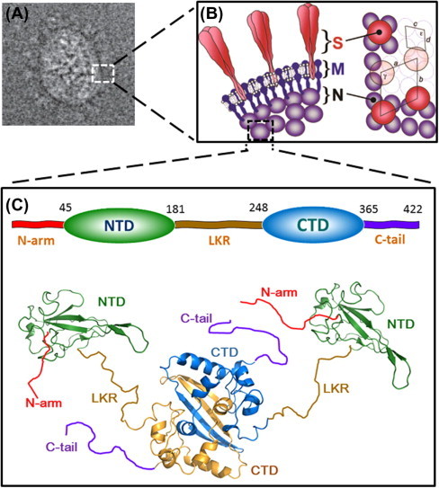
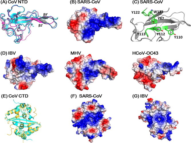
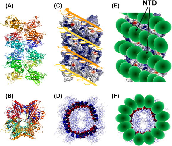
The nucleocapsid phosphoprotein of the severe acute respiratory syndrome coronavirus (SARS-CoV N protein) packages the viral genome into a helical ribonucleocapsid (RNP) and plays a fundamental role during viral self-assembly. It is a protein with multifarious activities. In this article we will review our current understanding of the N protein structure and its interaction with nucleic acid. Highlights of the progresses include uncovering the modular organization, determining the structures of the structural domains, realizing the roles of protein disorder in protein-protein and protein-nucleic acid interactions, and visualizing the ribonucleoprotein (RNP) structure inside the virions. It was also demonstrated that N-protein binds to nucleic acid at multiple sites with a coupled-allostery manner. We propose a SARS-CoV RNP model that conforms to existing data and bears resemblance to the existing RNP structures of RNA viruses. The model highlights the critical role of modular organization and intrinsic disorder of the N protein in the formation and functions of the dynamic RNP capsid in RNA viruses. This paper forms part of a symposium in Antiviral Research on "From SARS to MERS: 10 years of research on highly pathogenic human coronaviruses."

Keywords: Capsid packaging; Coronavirus; Intrinsic disorder; Nucleocapsid protein; RNP; SARS.


Figures

Fig. 1
Fig. 1
Structure of SARS-CoV N-protein. (A) 2D electron cryo-microscopy reconstructed image of SARS-CoV particle. (B) Interpretation of the virion structure. Edge view of the conserved structural proteins is shown on the left panel and the axial view is shown on the right panel. Trimeric spikes (S) are shaded in red, membrane proteins (M) are in solid blue, and nucleoproteins (N) are shaded in violet. The figures are reproduced with permission from Neuman et al. (2006). (C) The modular structural organization of SARS-CoV N protein. The domain boundaries shown on the top were defined by Chang et al. (2006a). The ribbon representations of the structures of NTD (green) and CTD (blue and gold) are generated with PyMOL from coordinates in the protein data bank (PDB IDs: NTD, 2OFX; CTD, 2CJR). The relative orientation of NTD and CTD, as well as the conformations of the disordered regions (N-arm, LKR and C-tail), are drawn randomly to reflect the dynamic nature of the N protein, as revealed by SAXS data (Chang et al., 2009). The ribbon structures were generated using PyMOL (The PyMOL Molecular Graphics System, Version 1.5.0.4 Schrödinger, LLC).
Fig. 2
Fig. 2
Multiple sequence alignments of coronavirus N proteins. Shaded positions represent conserved residues among the compared sequences. Residues in red denote aromatic residues that are postulated to be involved in base stacking interactions when binding to RNA. Secondary structure elements based on SARS-CoV N protein are shown on top of the alignment, with arrows and cylinders representing β-strands and α-helices, respectively. The alignment was calculated on the ClustalOmega server (http://www.ebi.ac.uk/Tools/msa/clustalo).
Fig. 3
Fig. 3
Structure comparisons of coronavirus N-proteins. (A) Structure comparison of various coronavirus NTDs (Grey: SARS-CoV, 2OFX; Magenta: IBV, 2GEC; Blue, MHV, 3HD4; Cyan: HCoV OC43. 4J3 K). The surface charge distributions on (B) (SARS-CoV) and (D) (IBV, MHV and HCoV OC43) are shown in same orientations. (C) Spatial arrangement of aromatic residues in NTD speculated to be involved in base stacking interaction when binding to RNA. Residues in the loop connecting β3 and β4 strands (a.a. Gly115-Gly130) have been removed for clarity. (E) Superimposition of the CTD structures of SARS CoV (gold, 2CJR) and IBV (cyan, 2GEC). The corresponding surface charge distributions were shown on (F) and (G) for SARS-CoV and IBV, respectively. All structures and surface charge distributions were generated using PyMOL.
Fig. 4
Fig. 4
A proposed model of the SARS-CoV ribonucleocapsid protein. The crystal packing of a 24-mer CTD domain is shown in side view (A) and top view (B). The surface charge distribution of the SARS-CoV CTD 24-mer. (C) Top view of the model shows the docking of two RNA chains (orange and yellow ribbons) onto the 24-mer CTD structure. The CTD 24-mer is shown in surface charge representation. The RNA chains were modeled with the phosphate backbone (red spheres) facing inside the groove and bases (yellow rings) pointing outward. (D) Top view of the putative CTD–RNA complex. (E) Schematic of the docking of NTD onto the CTD 24–mer-RNA complex. The NTD domains are represented by ellipsoids.

Comment in

References

    1. Ababou A., Ladbury J.E. Survey of the year 2005: literature on applications of isothermal titration calorimetry. J. Mol. Recognit. 2007;20:4–14. - PubMed
    1. Albertini A.A.V., Wernimont A.K., Muziol T., Ravelli R.B.G., Clapier C.R., Schoehn G., Weissenhorn W., Ruigrok R.W.H. Crystal structure of the rabies virus nucleoprotein–RNA complex. Science. 2006;313:360–363. - PubMed
    1. Ariza A., Tanner S.J., Walter C.T., Dent K.C., Shepherd D.A., Wu W.N., Matthews S.V., Hiscox J.A., Green T.J., Luo M., Elliott R.M., Fooks A.R., Ashcroft A.E., Stonehouse N.J., Ranson N.A., Barr J.N., Edwards T.A. Nucleocapsid protein structures from orthobunyaviruses reveal insight into ribonucleoprotein architecture and RNA polymerization. Nucleic Acids Res.2013;41:5912–5926. - PMC - PubMed
    1. Bárcena M., Oostergetel G.T., Bartelink W., Faas F.G.A., Verkleij A., Rottier P.J.M., Koster A.J., Bosch B.J. Cryo-electron tomography of mouse hepatitis virus: insights into the structure of the coronavirion. Proc. Nat. Acad. Sci. 2009;106:582–587. - PMC - PubMed
    1. Caul E.O., Egglestone S.I. Coronavirus-like particles present in simian faeces. Vet. Rec. 1979;104:168–169. - PubMed

Publication types

MeSH terms