Review
. 2014 Jan;70(Pt 1):2-20.
doi: 10.1107/S2053230X13033141. Epub 2013 Dec 24.

Introduction to protein crystallization

Affiliations
Review

Introduction to protein crystallization

Alexander McPherson et al. Acta Crystallogr F Struct Biol Commun. 2014 Jan.

Abstract

Protein crystallization was discovered by chance about 150 years ago and was developed in the late 19th century as a powerful purification tool and as a demonstration of chemical purity. The crystallization of proteins, nucleic acids and large biological complexes, such as viruses, depends on the creation of a solution that is supersaturated in the macromolecule but exhibits conditions that do not significantly perturb its natural state. Supersaturation is produced through the addition of mild precipitating agents such as neutral salts or polymers, and by the manipulation of various parameters that include temperature, ionic strength and pH. Also important in the crystallization process are factors that can affect the structural state of the macromolecule, such as metal ions, inhibitors, cofactors or other conventional small molecules. A variety of approaches have been developed that combine the spectrum of factors that effect and promote crystallization, and among the most widely used are vapor diffusion, dialysis, batch and liquid-liquid diffusion. Successes in macromolecular crystallization have multiplied rapidly in recent years owing to the advent of practical, easy-to-use screening kits and the application of laboratory robotics. A brief review will be given here of the most popular methods, some guiding principles and an overview of current technologies.

Keywords: IYCr; crystallization; proteins.


Figures
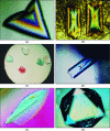
Figure 1
Figure 1
Microphotographs of protein and virus crystals grown in the laboratory of AM showing the variety of habits common to macromolecular crystals. (a, b, f) Satellite tobacco mosaic virus, (c) Desmodium yellow mottle virus, (d) hexagonal canavalin and (e) intact anti-canine lymphoma antibody.
Figure 2
Figure 2
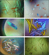
Shown here are a variety of protein crystals that were obtained directly from commercial screening matrices but, as is evident, some are inadequate for X-ray data collection because of morphology or size, implying that the crystallization conditions require optimization.
Figure 3
Figure 3
The phase diagram for the crystallization of macromolecules. The solubility diagram is divided sharply into a region of undersaturation and a region of supersaturation by the line denoting maximum solubility at specific concentrations of a precipitant, which may be salt or a polymer. The line represents the equilibrium between the existence of the solid phase and the free-molecule phase. The region of supersaturation is further divided in a more uncertain way into the metastable and labile regions. In the metastable region nuclei will develop into crystals, but no nucleation will occur. In the labile region both might be expected to occur. The final region, at very high supersaturation, is denoted the precipitation region, where this result might be most probable. Crystals can only be grown from a supersaturated solution, and creating such a solution supersaturated in the protein of interest is the immediate objective in growing protein crystals.
Figure 4
Figure 4
The sitting-drop vapor-diffusion method is illustrated in this schematic diagram. The drop on the elevated platform, which is commonly 2–10 µl, consists of half stock protein solution and half reservoir solution which contains some concentration of a salt or polymer precipitant. About 0.5 ml of the reservoir solution is added to the bottom of the cell before sealing. By water equilibration through the vapor phase the drop ultimately approaches the reservoir in osmolarity, both raising the concentration of the precipitant in the drop and increasing the protein concentration there.
Figure 5
Figure 5
The hanging-drop vapor-diffusion method is illustrated schematically. The components of the drop and reservoir, and the physical equilibration process, are the same here as for the sitting drop. The exception is that the protein drop is suspended from a cover slip over the reservoir rather than resting on a surface. Plasticware for carrying out both sitting- and hanging-drop vapor diffusion are widely and commercially available in numerous formats.
Figure 6
Figure 6
The use of microdialysis buttons to dialyze small volumes of protein solution against a precipitating solution is illustrated. The protein solution volumes may be from 10 to 50 µl. The buttons are commercially available.
Figure 7
Figure 7
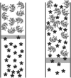
The process of free-interface diffusion to effect crystallization is illustrated. A protein solution is layered atop a precipitant solution in a narrow-bore tube or capillary. Diffusion across the interface, principally of the precipitant, induces nucleation and growth.
Figure 8
Figure 8
Diagram illustrating the counter-diffusion method for growing protein crystals. Here, the protein solution is shown in red and the gel saturated with the precipitant solution is shown in green. The capillaries are sealed at their distal end but are open where they enter the gel. By diffusion of precipitant up the length of the capillary, a concentration gradient is formed that explores a wide range of precipitant conditions.
Figure 9
Figure 9
The curve shown here represents a typical solubility curve for a protein and divides, as in Fig. 2 ▶, the region of undersaturation from that of supersaturation. It also illustrates the existence of the classical ‘salting-in’ and ‘salting-out’ regions for the protein. By taking advantage of these latter effects, supersaturation may be achieved by equilibrating a system from a point of maximum solubility (P 0) to one of reduced solubility (P 1 or P 2) by adjusting the precipitant concentration.
Figure 10
Figure 10
This diagram, based on an analysis of nearly 2800 examples, shows the distribution of the number of protein crystals grown as a function of pH. As one might expect, the great majority have been grown near neutrality, reflecting the desire of investigators to crystallize their protein near physiological conditions. The spread, however, illustrates that protein crystals might reasonably be expected over a very large pH range and that this entire range deserves attention (figure courtesy of Hampton Research, Aliso Viejo, CA, USA).
Figure 11
Figure 11
As shown here, most proteins have certain solubility minima as a function of pH. One can take advantage of this property to produce supersaturation by altering a system from a pH permitting high solubility (P 1 or P 2) to a point of low solubility (P 0). This is a powerful approach to promoting crystallization of macromolecules.
Figure 12
Figure 12
An observation of inadvertent heterogeneous nucleation of protein crystals that is not uncommon is that of crystals growing along the length of a cotton fiber present in the mother liquor. These are crystals of a Fab fragment from an IgG.
Figure 13
Figure 13
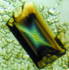
Shown here is a single large crystal of Satellite tobacco mosaic virus that is approximately 1.5 mm in the longest dimension and which shows a high degree of birefringence under polarized light. This crystal was grown in microgravity aboard the US Space Shuttle in 1991.

Similar articles

Cited by

References

    1. Abel, J. J., Geiling, E. M. K., Roultier, O. A., Bell, F. M. & Wintersteiner, O. (1927). Crystalline insulin. J. Pharmacol. Exp. Ther. 31, 65–85.
    1. Bailey, K. (1942). The growth of large single crystals of proteins. Faraday Soc. Trans. 38, 186–192.
    1. Bard, J., Ercolani, K., Svenson, K., Olland, A. & Somers, W. (2004). Automated systems for protein crystallization. Methods, 34, 329–347. - PubMed
    1. Bergfors, T. M. (1999). Protein Crystallization: Techniques, Strategies, and Tips La Jolla: International University Line.
    1. Bergfors, T. (2003). Seeds to crystals. J. Struct. Biol. 142, 66–76. - PubMed