

Purification, crystallization and preliminary X-ray diffraction analysis of a novel keto-deoxy-D-galactarate (KDG) dehydratase from Agrobacterium tumefaciens
- PMID: 24419616
- PMCID: PMC3943101
- DOI: 10.1107/S2053230X13031361
Purification, crystallization and preliminary X-ray diffraction analysis of a novel keto-deoxy-D-galactarate (KDG) dehydratase from Agrobacterium tumefaciens
Abstract
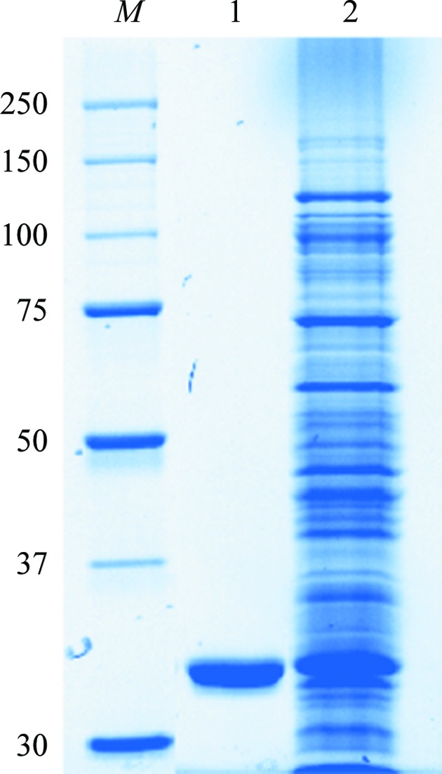
D-galacturonic acid is the main component of pectin. It could be used to produce affordable renewable fuels, chemicals and materials through biotechnical conversion. Keto-deoxy-D-galactarate (KDG) dehydratase is an enzyme in the oxidative pathway of D-galacturonic acid in Agrobacterium tumefaciens (At). It converts 3-deoxy-2-keto-L-threo-hexarate to α-ketoglutaric semialdehyde. At KDG dehydratase was crystallized by the hanging-drop vapour-diffusion method. The crystals belonged to the monoclinic space group C2, with unit-cell parameters a = 169.1, b = 117.8, c = 74.3 Å, β = 112.4° and an asymmetric unit of four monomers. X-ray diffraction data were collected to 1.9 Å resolution using synchrotron radiation. The three-dimensional structure of At KDG dehydratase will provide valuable information on the function of the enzyme and will allow it to be engineered for biorefinery-based applications.
Keywords: Agrobacterium tumefaciens; d-galacturonic acid; keto-deoxy-d-galactarate dehydratase; oxidative pathway.
Figures

References
Publication types
MeSH terms
Substances
LinkOut - more resources
Full Text Sources
Other Literature Sources
Miscellaneous
