Review
. 2013:54:65-116.
doi: 10.1007/978-3-642-41004-8_4.

Polyoxometalates active against tumors, viruses, and bacteria

Affiliations
Review

Polyoxometalates active against tumors, viruses, and bacteria

Toshihiro Yamase. Prog Mol Subcell Biol. 2013.

Abstract

Polyoxometalates (PMs) as discrete metal-oxide cluster anions with high solubility in water and photochemically and electrochemically active property have a wide variety of structures not only in molecular size from sub-nano to sub-micrometers with a various combination of metals but also in symmetry and highly negative charge. One of the reasons for such a structural variety originates from their conformation change (due to the condensed aggregation and the structural assembly) which strongly depends on environmental parameters such as solution pH, concentration, and coexistent foreign inorganic and/or organic substances. In the course of the application of the physicochemical properties of such PMs to the medical fields, antitumoral, antiviral, and antibacterial activities have been developed for realization of a novel inorganic medicine which provides a biologically excellent activity never replaced by other approved medicines. Several PMs as a candidate for clinical uses have been licensed toward the chemotherapy of solid tumors (such as human gastric cancer and pancreatic cancer), DNA and RNA viruses (such as HSV, HIV, influenza, and SARS), and drug-resistant bacteria (such as MRSA and VRSA) in recent years: [NH3Pr(i)]6[Mo7O24]∙3H2O (PM-8) and [Me3NH]6[H2Mo(V) 12O28(OH)12(Mo(VI)O3)4]∙2H2O (PM-17) for solid tumors; K7[PTi2W10O40]∙6H2O (PM-19), [Pr(i)NH3]6H[PTi2W10O38(O2)2]∙H2O (PM-523), and K11H[(VO)3(SbW9O33)2]∙27H2O (PM-1002) for viruses; and K6[P2W18O62]∙14H2O (PM-27), K4[SiMo12O40]∙3H2O (SiMo12), and PM-19 for MRSA and VRSA. The results are discussed from a point of view of the chemotherapeutic clarification in this review.


Figures
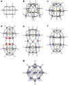
Fig. 4.1
Fig. 4.1
Structures depicted by atom and bond model for anions of some of the bioactive PMs. [MoVI 7O24]6− (a), [H2MoV 12O28(OH)12(MoVIO3)4]6− (b), [PTi2W10O40]7− (c), [(GeTi3W9O37)2O3]14− (d), [(VIVO)3(SbW9O33)2]12− (e), [VIV 18O42(Cl)]13− (f), and [P2W18O62]6− (g). (a) and (b) correspond to the anion for antitumoral [NH3Pri]6[Mo7O24]∙3H2O (PM-8) and [Me3NH]6[H2MoV 12O28(OH)12(MoVIO3)4]∙2H2O (PM-17), respectively, in which MoVI, MoV, and O atoms are indicated by blue, brown, and white color, respectively. (c), (d), (e), and (f) correspond to the anion for antiviral K7[PTi2W10O40]∙6H2O (PM-19), K9H5[(GeTi3W9O37)2O3]∙16H2O (PM-504), K11H[(VO)3(SbW9O33)2]∙27H2O (PM-1002), and K6[P2W18O62]∙14H2O (PM-27), respectively, in which WVI, TiIV (or VIV), PV (GeIV or SbIII), and O atoms are indicated by blue, red, yellow, and white color, respectively. (g) Corresponds to the anion for a strong sialyl/sulfotransferase inhibitor, K10H3[V18O42(Cl)]∙12H2O, which is also antibacterial against Streptococcus pneumoniae. In (f) VIV, Cl, and O atoms are indicated by blue, red, and white color, respectively
Fig. 4.2
Fig. 4.2
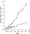
Inhibition of tumor growth by PM-8 and PM-17 after implantation of MX-1. PM-8 (open circles) was i.p. administrated by 200 mg/kg/day on days 17–27 except 19 and 100 mg/kg/day on days 17, 18, and 20, and PM-17 (open triangles) was done by 25 mg/kg/day on days 21–27. Control (filled circles) showed no PM treatment. The change in body weights on days 17–32 for PM-17-treated mice was −5.0 g, while there was no significant change on days 17–46 for PM-8-treated mice
Fig. 4.3
Fig. 4.3
Contents of Mo atoms in the liver, kidney, brain, plasma, and tumor for the tumor mice at 0.5 h after i.p. administration of PM-8, which were determined by the radioactivation (to 101Mo) analysis. The contents for PM-8 untreated mice (both tumor mice and normal mice) as backgrounds are also indicated for comparison
Fig. 4.4
Fig. 4.4
Time profile of the Mo contents in the kidney and tumor for the tumor mice after i.p. administration of PM-8
Fig. 4.5
Fig. 4.5
Differential pulse polarograms of FMN (2 mM), PM-8 (2 mM), and their mixture in aqueous solutions (at pH 5) containing 0.1 M NaClO4, which are depicted by blue, green, and red curves, respectively. DC polarogram of the mixture is shown by black line for comparison. Top figure shows ESR spectrum observed by the one-electron reduction of the mixture
Fig. 4.6
Fig. 4.6
Effect of PM-17 on in vitro survival of AsPC-1 (a) and MKN-45 (b) cells (1.5 × 105 cells/5 ml for each cell) for 24 h. Each point indicates the percentage of living cell in three independent experiments (bars depict s.d.)
Fig. 4.7
Fig. 4.7
In vivo tumor growth inhibition by PM-17 for AsPC-1 implanted mice. 2 × 106 AsPC-1 cells were transplanted into the back of Balb/c nude mice, and after 10 days, intratumoral injections of PM-17 (125 μg or 500 μg dissolved in 100 μl of saline) were performed for 10 days with 2-day intermission on day 6. The control mice were treated with 100 μl of saline per day under the same conditions. Changes in tumor volume (a) and body weight (c) for five mice per group are depicted with photographs (b) for the mice on day 41after the tumor implantation
Fig. 4.8
Fig. 4.8
Hematoxylin and eosin (H&E) (a) and terminal deoxynucleotidyl transferase-mediated “nick-end” labeling (TUNEL) (b) stainings for thin sections of the tumor on the 10th day after a single intratumoral injection of PM-17 (500 μg/100 μl saline) into the AsPC-1 transplanted nude mice, in comparison with the control (with the injection of 100 μl 0.9 % NaCl saline)
Fig. 4.9
Fig. 4.9
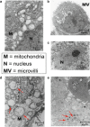
TEM images of the AsPC-1 cells treated with (ce) and without (a and b) PM-17
Fig. 4.10
Fig. 4.10
In vitro expression of autophagy by the green fluorescent protein (GFP)-tagged light-chain 3 (LC3) in the PM-17-treated AsPC-1 cells. 2 × 105 AsPC-1 cells were cultured with the BRMI-1640 growth medium for 24 h in 24-wells plate, transfected with GFP-LC3 expression plasmid (0.8 μg/well) with lipofectamine 2000 transfection reagent (2 μg/well). After 24 h the cells were treated with and without PM-17 (175 μg/ml saline) for 24 h, and the distribution (a) of fluorescence of GFP-LC3 under fluorescence microscope and a Western blotting picture (b) were investigated
Fig. 4.11
Fig. 4.11
In vivo cytotoxicity (a) and antiviral activity (b) for three PMs, 523, 1,002, and 1,208. Survival time indicates the time (in h) after i.p. administration of PM of 125 (open square), 250 (filled square), 500 (open circle), and 1,000 (filled circle) mg/kg/mouse. The activity of the infected virus (vPE 16, vP 1,206, or WR) in mice was evaluated on day 5, by the β-galactosidase activity for ovaries of mice which were i.p. administrated by 12.5 and 125 mg/kg/day/mouse of each PM (PM -523, PM -1002, or PM -1208) for 3 days from days 2 to 4 after the virus infection on day 0
Fig. 4.12
Fig. 4.12
Synergistic therapeutic effect in mice infected with FluV-A (H1N1/PR8) using PM-523 and ribavirin. All of the control mice (inbred 8-week-old female Balb/c) were infected with ten lethal doses of FluV-A, which corresponds to 1.6 × 104 median tissue culture infection dose (TCID50) titers in MDCK cell line. From 8 h after infection, the mice (ten mice/group) were treated with aerosols of PM-523 and ribavirin either singly or in combination every 12 h for 4 days. Compound solutions were prepared in phosphate-buffered saline (PBS) solutions (at pH 7.2), and the infected mice were exposed to the compounds by using a continuous aerosol generator for 2 h at a rate of distribution of 120 μl of solution/h to each chamber. Each infected 20-g mouse was placed in a chamber separately; no compound (filled circle)-treated, 40 mM ribavirin (filled triangle)-treated, 80 mM ribavirin (filled square)-treated, 2.4 mM PM-523 (open triangle)-treated, 4.8 mM PM-523 (open square)-treated, and 40 mM ribavirin/2.4 mM PM-523 combination (open circle)-treated mice. Levels of significance of p < 0.001 for 2.4-mMPM-523/40-mM ribavirin combination, p < 0.01 for 4.8-mM PM-523, and p < 0.05 for 80-mM ribavirin-treated mice on day 14 after infection
Fig. 4.13
Fig. 4.13
Change of viral titers in the lungs of the infected mice with and without treatment of PM-523 and/or ribavirin after infection (1.6 × 104 TCID50 of FluV/A/PR8/mouse). The values for the uninfected and untreated mice are plotted for comparison. Uninfected and untreated (open triangle), infected and untreated (filled circle), 40-mM ribavirin-treated (filled square), 2.4-mMPM-523-treated (open square), and 40 mM ribavirin/2.4 mM PM-523 combination-treated (open circle) mice
Fig. 4.14
Fig. 4.14
Inhibition mode of PM-504 on the enzymatic activity of ST3Gal-1 at various concentrations of core 1 (a) and CMP-Neu5Ac (b) in the absence and presence of PM-504. (a): 0 (filled circle), 0.15 nM (open circle), and 0.3 nM (filled square) of PM-504 in the presence of 1.6-μM CMP-Neu5Ac. (b): 0 (filled circle), 0.1 nM (open circle), and 0.2 nM (filled square) of PM-504 in the presence of 1-mM core 1
Fig. 4.15
Fig. 4.15
Growth-inhibitory zone around the disk for the Mu50 strain treated with PM-19 at concentrations of 50 (a), 100 (b), and 500 (c) μM
Fig. 4.16
Fig. 4.16
Effects of PM alone (a) and the coexistence of PM and oxacillin (b) on the electrophoresis results of the RT-PCR products for MRSA SR3605. SR3605 strain cells were incubated in Muller–Hinton broth in the presence (a) of PM (PM-27, SiMo12, or PM-19) at various concentrations indicated by MIC units or in the coexistence (b) of PM (at various concentrations less than MIC) and oxacillin (at 1/4 MIC). Lane M indicates 100 bp DNA ladder as molecular weight marker
Fig. 4.17
Fig. 4.17
Effects of PM alone (a) and the coexistence of PM and oxacillin (b) on the electrophoresis results of the RT-PCR products for VRSA Mu50. Mu50 strain cells were incubated in MH broth in the presence (a) of PM (PM-27, SiMo12, or PM-19) at various concentrations indicated by MIC units or in the coexistence (b) of PM (at various concentrations less than MIC) and oxacillin (at 1/4 MIC). Lane M indicates 100 bp DNA ladder as molecular weight marker
Fig. 4.18
Fig. 4.18
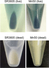
Blue coloration of live cells of both SR3605 and Mu50 treated with PM-27. Photographs indicate the centrifuged cells of SR3605 or Mu50 strain in saline suspension containing 1 mM PM-27. Dead cells were prepared by the treatment of the cells with ethanol for 1 day
Fig. 4.19
Fig. 4.19
TEM images of the MRS396-1 cells treated with and without PM-19 and elemental spectra of local points of the cells (A–D for PM-treated and A and B for untreated ones). Inoculum (108 cfu, colony-forming unit/ml) of MRS394-1 was inoculated in MH broth containing 40 mM PM-27 for 5 h at 34 °C. The cells harvested by centrifugation (with 5,000 × g for 15 min at 4 °C) and washed twice with PBS were fixed with 2.5 % glutaraldehyde in PBS and followed by dehydration with a graded series of ethanol, embedding into a resin, and sectioning by conventional method. The spectrum observed on JEOL JEM-2100 F was examined by the distribution of W and Ti atoms in the cells with a help of energy dispersive X-ray analysis (JEOL JED-2300 T)
Fig. 4.20
Fig. 4.20
Amounts of W atoms uptaken in the cells for the bacteria cells of SR3605 (a) and Mu50 (b) strains, cultured with 1/4 MIC oxacillin, 1/4 PM-19, or a combination of PM-19 and oxacillin (1/4 MIC for each). **p < 0.01 for n = 3. Compound-untreated cells (control) showed no observable amount of W atoms in the cells
Fig. 4.21
Fig. 4.21
Amounts of W atoms (of PM-27) uptaken into the Mu50 cells for six cultivation modes of the Mu50 cells. Six cultivation modes are (1) cultivation without substrate (control), (2) 1/4 MIC oxacillin alone until the stage of a logarithmic growth phase (OD660 ≈ 0.2), (3) 1/4 MIC PM-27 alone until the stage of a logarithmic growth phase (OD660 ≈ 0.2), (4) at first 1/4 MIC oxacillin until the logarithmic growth phase and subsequent 1/4 MIC PM-27 for 24 h, (5) at first 1/4 MIC PM-27 until the logarithmic growth phase and subsequent 1/4 MIC oxacillin for 24 h, and (6) a combination of PM-19 and oxacillin (1/4 MIC for each) until the stage of a logarithmic growth phase (OD660 ≈ 0.2). **p < 0.01 for n = 3

Similar articles

Cited by

References

    1. Baba M, Snoeck R, Pauwels R, DeClerq E. Sulfated polysaccharides are potent and selective inhibitors of various enveloped viruses including herpes simplex viruses, cytomegalovirus, vesicular stomatitis viruses, and human immunodeficiency virus. Antimicrob Agents Chemother. 1988;32:1742–1745. - PMC - PubMed
    1. Barnard DL, Hill CL, Gage T, Matheson JE, Huffmann JH, Sidwell RW, Otto MI, Schinazi RF. Potent inhibition of respiratory syncytial virus by polyoxometalates of several structure classes. Antiviral Res. 1997;34:27–37. - PubMed
    1. Blumenthal R, Clgue MJ, Durell SR, Epand RM. Membrane fusion. Chem Rev. 2003;103:53–69. - PubMed
    1. Boneca IG, Chiosis G. Vancomycin resistance: occurrence, mechanism and strategies to combat it. Expert Opin Ther Targets. 2003;7:311–328. - PubMed
    1. Borrás-Alamenar JJ, Coronado E, Müller A, Pope MT, editors. Polyoxometalate molecular science. Dordrecht: Kluwer; 2003.

Publication types

MeSH terms