. 2014 Apr 24;123(17):2703-14.
doi: 10.1182/blood-2013-07-516948. Epub 2014 Jan 13.

The F-BAR protein PSTPIP1 controls extracellular matrix degradation and filopodia formation in macrophages

Affiliations

The F-BAR protein PSTPIP1 controls extracellular matrix degradation and filopodia formation in macrophages

Taylor W Starnes et al. Blood. .

Abstract

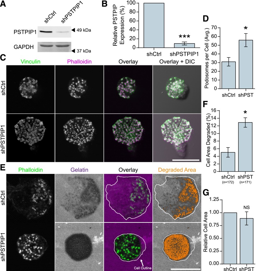
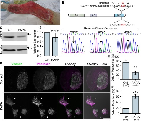
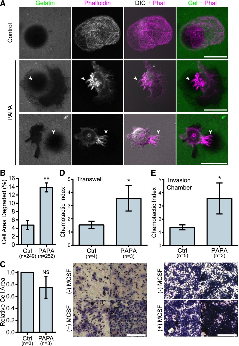
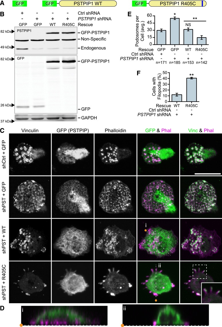
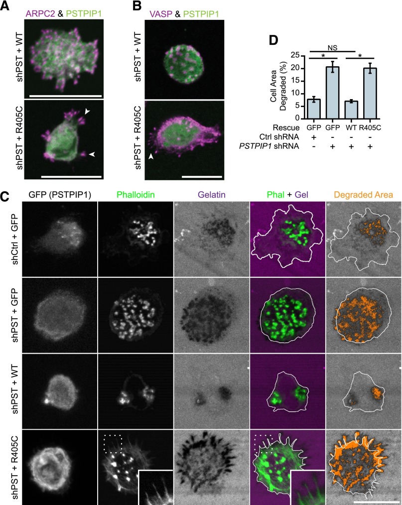
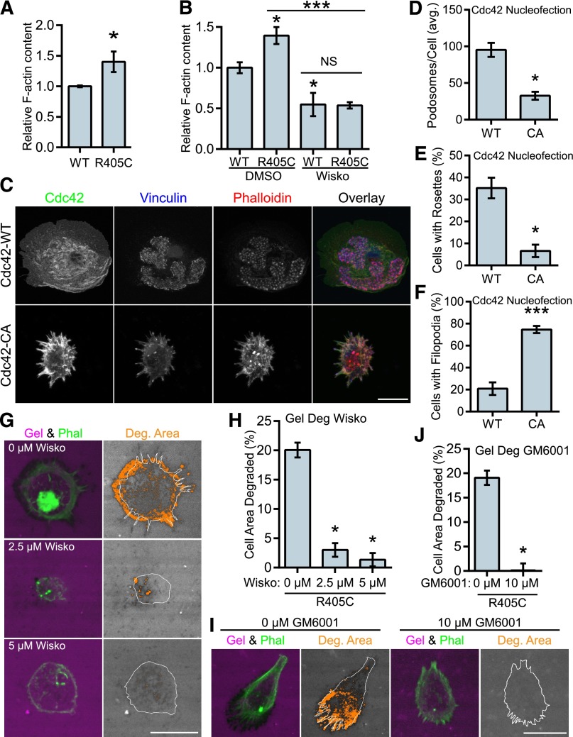
PSTPIP1 is a cytoskeletal adaptor and F-BAR protein that has been implicated in autoinflammatory disease, most notably in the PAPA syndrome: pyogenic sterile arthritis, pyoderma gangrenosum, and acne. However, the mechanism by which PSTPIP1 regulates the actin cytoskeleton and contributes to disease pathogenesis remains elusive. Here, we show that endogenous PSTPIP1 negatively regulates macrophage podosome organization and matrix degradation. We identify a novel PSTPIP1-R405C mutation in a patient presenting with aggressive pyoderma gangrenosum. Identification of this mutation reveals that PSTPIP1 regulates the balance of podosomes and filopodia in macrophages. The PSTPIP1-R405C mutation is in the SRC homology 3 (SH3) domain and impairs Wiskott-Aldrich syndrome protein (WASP) binding, but it does not affect interaction with protein-tyrosine phosphatase (PTP)-PEST. Accordingly, WASP inhibition reverses the elevated F-actin content, filopodia formation, and matrix degradation induced by PSTPIP1-R405C. Our results uncover a novel role for PSTPIP1 and WASP in orchestrating different types of actin-based protrusions. Our findings implicate the cytoskeletal regulatory functions of PSTPIP1 in the pathogenesis of pyoderma gangrenosum and suggest that the cytoskeleton is a rational target for therapeutic intervention in autoinflammatory disease.


Figures

Figure 1
Figure 1
PSTPIP1 impairs podosome formation. (A-B) Lentiviral transduction of shRNA was used to knock down PSTPIP1 in THP-1 cells. (A) Western blot of lysates from the control (shCtrl) and PSTPIP1 knockdown (shPSTPIP1 or shPST) THP-1 cells using anti-PSTPIP1 and anti–glyceraldehyde 3-phosphate dehydrogenase (GAPDH) antibodies (loading control). Arrowheads indicate location of molecular weight markers. (B) Quantification of PSTPIP1 knockdown in which PSTPIP1 expression in shCtrl cells was normalized to 100%. (C) Representative images of podosomes in shCtrl and shPSTPIP1 THP-1 macrophages. Podosomes were stained with anti-vinculin antibody (green) and rhodamine phalloidin (magenta). (D) Quantification of the average number of podosomes formed by shCtrl and shPSTPIP1 THP-1 cells. (E) Representative images of gelatin degradation by PSTPIP1 knockdown cells on Alexa Fluor 568–conjugated gelatin (magenta) coverslips. Podosomes were stained with Alexa Fluor 488 phalloidin (green). The cell outline is a trace of the cell border made from the phalloidin image. Orange pixels in the Degraded Area panel were those counted as degraded. (F) Quantification of the amount of gelatin degraded by PSTPIP1 knockdown cells as a percentage of cell area. (G) Cell area was determined in all cells for the degradation experiment and normalized to the shCtrl value. Scale bars, 20 μm. All values are mean ± standard error of the mean (SEM) from 3 (D,F,G) or 4 (B) independent experiments. *P < .05; ***P < .001 as determined by a paired Student t test (B,D,G) or ANOVA with repeated measures using a compound symmetry correlation structure (F). NS, nonsignificant.
Figure 2
Figure 2
Macrophages from a PAPA syndrome patient have increased filopodia-like membrane projections. (A) Image of pyoderma gangrenosum on the PAPA patient’s right leg. (B) Reverse strand sequence traces from the PSTPIP1 genomic DNA sequencing of the patient, his mother, and his father. The diagram shows the location of the mutation at the DNA and protein level. (C) Western blots of lysate from control and PAPA macrophages was probed with mouse anti-PSTPIP1 and rabbit anti-β-tubulin (loading control) antibodies to determine relative PSTPIP1 expression. Arrowheads indicate location of molecular weight markers (kDa). PSTPIP1 expression in the PAPA patient macrophages was normalized to controls for quantification. (D) Representative images of podosomes and filopodia-like membrane projections in PAPA and control macrophages. Podosomes and filopodia-like structures were stained with anti-vinculin antibody (green) and rhodamine phalloidin (magenta). Arrowheads indicate filopodia-like projections in PAPA macrophages. (E-F) Quantification of the percentage of macrophages shown in (D) that form podosomes (E) and filopodia-like projections (F). Scale bar, 20 μm. All values are mean ± SEM from 3 (E,F) or 4 (C) independent experiments. **P < .01; ***P < .001 as determined by an unpaired Student t test (E,F) or paired Student t test (C).
Figure 3
Figure 3
PAPA patient macrophages make membrane projections associated with matrix degradation and have enhanced invasive properties. (A) Representative images of gelatin degradation by control and PAPA macrophages on Oregon Green 488 gelatin-coated coverslips (green). Podosomes and membrane projections were stained with rhodamine phalloidin (magenta). Areas degraded by the macrophages appear black in the gelatin image. Arrowheads indicate projections overlaying degraded areas. (B) Quantification of gelatin degraded by control and PAPA macrophages as a percentage of the total cell area. (C) Cell area was determined for all cells in the assay and normalized to the value of the control macrophages. (D-E) Quantification of macrophage chemotaxis to macrophage colony-stimulating factor (M-CSF) in (D) Transwell devices or (E) Matrigel-coated invasion chambers. Chemotactic index is the ratio of cells migrating to M-CSF over medium alone. Representative images of the filters are shown below. Scale bars, 20 μm (A) or 500 μm (D,E). All values are mean ± SEM from 3 independent experiments. *P < .05; **P < .01 as determined by a paired Student t test (C) unpaired Student t test (D,E) or ANOVA with repeated measures using a compound symmetry correlation structure (B).
Figure 4
Figure 4
PSTPIP1-R405C promotes filopodia formation. (A) Diagram of constructs introduced into control and PSTPIP1 knockdown cells by retroviral transduction to make rescue cell lines. (B) Representative western blot of lysates from FACS-sorted THP-1 rescue lines showing expression of rescue constructs. Blots were probed with mouse anti-PSTPIP1, rabbit anti-GFP, and mouse anti-GAPDH (loading control) antibodies. Arrowheads indicate location of molecular weight markers. (C) Representative images of podosomes and filopodia formed by the THP-1 rescue cell lines, which were stained with anti-vinculin antibody (green) and rhodamine phalloidin (magenta). Anti-GFP antibody (green) was used to amplify the GFP signal. Inset shows filopodia at ×1.5 original magnification. (D) Slices through the Z-plane of the shPST + WT and shPST + R405C cells were created with the Reslice function in FIJI software as indicated by the dashed lines in (C). (E) Quantification of the average number of podosomes formed by the indicated rescue line. shCtrl + GFP was the reference value for statistical comparison except where indicated by a bar. (F) Quantification of the percentage of shPST + WT and shPST + R405C cells that form filopodia. Scale bars, 20 μm. All values are mean ± SEM from 3 independent experiments. *P < .05; **P < .01 as determined by ANOVA with repeated measures using a compound symmetry correlation structure (E) or paired Student t test (F). NS, nonsignificant.
Figure 5
Figure 5
Filopodia associated with matrix degradation are induced by PSTPIP1 R405C. (A-B) Representative images of podosomes and filopodia formed in the THP-1 rescue cell lines. (A) The Arp2/3 complex was stained in podosomes and filopodia using anti-ARPC2 antibody (magenta) in THP-1 rescue cells expressing GFP-tagged PSTPIP1 constructs (green). (B) VASP was labeled in podosomes and filopodia with anti-VASP antibody (magenta) in THP-1 rescue cells expressing GFP-tagged PSTPIP1 constructs (green). (C) Representative images of gelatin degradation in the THP-1 rescue lines plated on Alexa Fluor 568–conjugated gelatin-coated coverslips (magenta). F-actin was stained with CruzFluor 405 phalloidin (green), and anti-GFP antibody (gray) was used to amplify the GFP signal. Orange pixels in the Degraded Area panel were those counted as degraded. (D) Quantification of the amount of gelatin degraded by the THP-1 rescue cells as a percentage of cell area. Scale bar, 20 μm. Values are mean ± SEM from 4 independent experiments. *P < .05, as determined by one-way ANOVA with Sidak’s multiple comparisons test. NS, nonsignificant.
Figure 6
Figure 6
PSTPIP1-R405C has impaired interaction with WASP but not PTP-PEST. (A) Anti-GFP antibody was used to immunoprecipitate GFP or GFP-tagged PSTPIP1 constructs from the indicated THP-1 rescue cell lines. Blots were probed with anti-GFP antibody and anti-WASP antibody to visualize the coimmunoprecipitation (IP) of WASP. Equivalent loading was determined by probing a blot of the lysates as above. (B) His-tagged WASP or GST-tagged PSTPIP1 proteins (labeled as GST-WT, GST-R405C, and GST-ΔSH3) were purified from E coli. They were separated by sodium dodecyl sulfate polyacrylamide gel electrophoresis (SDS-PAGE) and stained with Coomassie brilliant blue to assess purity. (C) Pulldown assay using the purified proteins described in (B). Soluble His-WASP was incubated with the indicated bead-bound GST-PSTPIP1 construct. Blots were probed for WASP and GST (to detect PSTPIP1) to determine the degree of interaction. The amount of WASP pulled down by the GST-PSTPIP1 mutants was normalized to the interaction with GST-PSTPIP1-WT. (D) Anti-GFP antibody was used to coimmunoprecipitate PTP-PEST with GFP or GFP-tagged PSTPIP1 constructs from the indicated THP-1 rescue cell lines. Anti–PTP-PEST and anti-GFP antibodies were used to probe the immunoprecipitation and input blots. (E) Quantification of the amount of PTP-PEST coimmunoprecipitating with PSTPIP1 was determined as a percent of input and normalized to the PSTPIP1-WT value. (F) Anti-GFP antibody was used to immunoprecipitate GFP-PSTPIP1-WT or -R405C from THP-1 rescue cells. Immunoprecipitates and lysates were probed with anti-phosphotyrosine (4G10 platinum) and anti-GFP antibodies to assess PSTPIP1 phosphorylation. (G) Quantification of the phosphorylation of PSTPIP1-WT and -R405C is shown relative to WT. Unlabeled arrowheads indicate the band(s) of interest in each panel. Labeled arrowheads indicate the locations of the molecular weight markers (kDa). All values are mean ± SEM from 3 (C,G) or 4 (E) independent experiments. **P < .01; ****P < .0001 as determined by one-way ANOVA with Sidak’s multiple comparisons test (C,E) or paired Student t test (G). IB, immunoblotting. NS, nonsignificant.
Figure 7
Figure 7
WASP mediates the transition to filopodia in PSTPIP1-R405C cells. (A) The F-actin polymerizing ability of PSTPIP1-WT and -R405C rescue cells was determined by stimulating serum-starved cells with chemokine (C-C motif) ligand 2. F-actin was saturated with rhodamine phalloidin and extracted with methanol; fluorescence was determined with a plate reader. Fluorescence intensity was normalized to the level of PSTPIP1-WT cells for each experiment. (B) F-actin content assay performed as in (A). Prior to stimulation, cells were pretreated for 30 minutes with dimethyl sulfoxide (DMSO) or 10 μM wiskostatin. Stimulation was performed in the presence of DMSO or 20 μM wiskostatin (Wisko) for 5 minutes. DMSO-treated WT was the reference value for statistical comparison except where indicated by a bar. (C-F) Macrophages nucleofected with GFP-Cdc42 WT (WT) or GFP-Cdc42 Q61L (CA) were plated on fibrinogen-coated coverslips overnight and stained with rhodamine phalloidin, anti-vinculin antibody, and anti-GFP antibody to visualize podosomes and Cdc42 expression. (C) Representative images of podosomes and filopodia in Cdc42-nucleofected cells. GFP-positive cells were used for (D) quantification of the average number of podosomes per cell, (E) the percentage of cells that made podosome rosettes, and (F) the percentage of cells forming filopodia. (G-J) THP-1 shPST + R405C rescue cells were plated on Alexa Fluor 568–conjugated gelatin for 3 hours with the indicated concentration of (G) wiskostatin or (I) GM6001. Representative images are shown. (H) The amount of degradation in the presence of wiskostatin is shown as a percentage of cell area (0 μM, n = 112; 2.5 μM, n = 134; 5 μM, n = 138). (J) The amount of degradation in the presence of GM6001 is shown as a percentage of cell area (0 μM, n = 125; 10 μM, n = 131). All values are mean ± SEM from 3 (B,H,J) or 4 (A,D-F) independent experiments. *P < .05; ***P < .001 as determined by (A) unpaired Student t test, (B) one-way ANOVA with Sidak’s multiple comparison test, (D-F) paired Student t test, or (H,J) ANOVA with repeated measures using a compound symmetry correlation structure.

Comment in

References

    1. Badolato R. Defects of leukocyte migration in primary immunodeficiencies. Eur J Immunol. 2013;43(6):1436–1440. - PubMed
    1. Friedl P, Weigelin B. Interstitial leukocyte migration and immune function. Nat Immunol. 2008;9(9):960–969. - PubMed
    1. Mackay CR. Moving targets: cell migration inhibitors as new anti-inflammatory therapies. Nat Immunol. 2008;9(9):988–998. - PubMed
    1. Lokuta MA, Cooper KM, Aksentijevich I, Kastner DL, Huttenlocher A. Neutrophil chemotaxis in a patient with neonatal-onset multisystem inflammatory disease and Muckle-Wells syndrome. Ann Allergy Asthma Immunol. 2005;95(4):394–399. - PubMed
    1. Heath RJW, Insall RH. F-BAR domains: multifunctional regulators of membrane curvature. J Cell Sci. 2008;121(Pt 12):1951–1954. - PubMed

Publication types

MeSH terms

Supplementary concepts